ANATOMY & PHYSIOLOGY · SECTION 7.2
Inside a Long Bone
Follow along with your worksheet, one question at a time.
mrsajdak.com/longbone
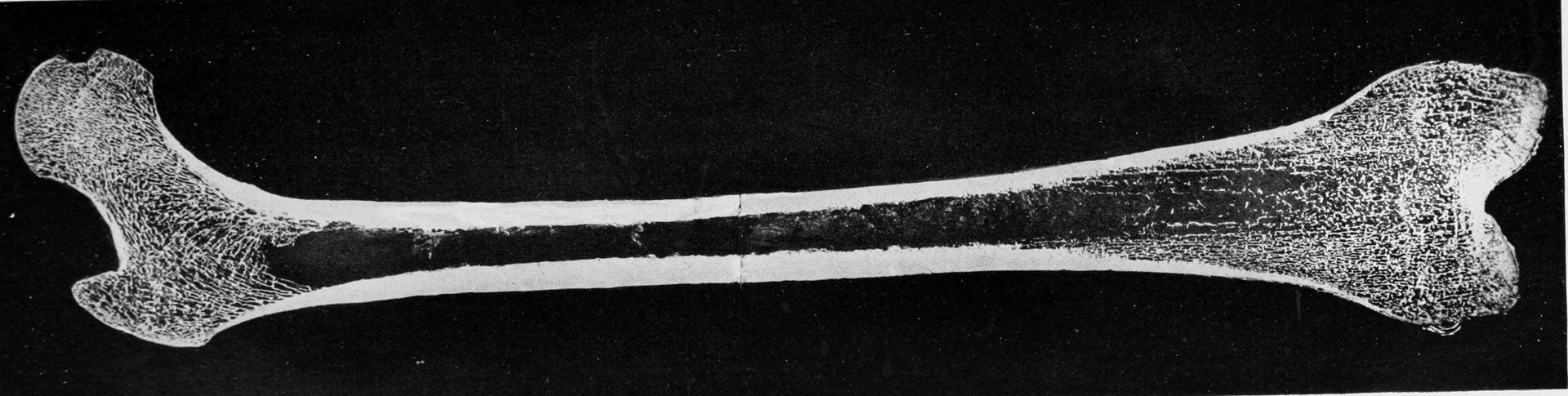
Photo: The American Journal of Anatomy (1917), public domain
START HERE · PICK AN OPENER
Choose how to open the lesson
Click a card to jump to it. Every option ends at the same Observe, Wonder, Predict slide and the same driving question.
OPTION A · VIDEO
A Needle Into Bone
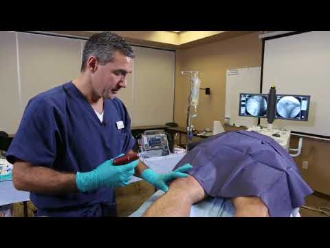
Clip 1 is a training film from the maker of a needle driver. It shows where the needle goes on the shin bone. Clip 2 is a short explainer of the same idea.
Watch for
Where on the leg does the needle go in?
Why would anyone put a needle in a bone?
What do you think is at the tip of the needle?
Videos on YouTube: Teleflex; YouTube.
OPTION B · TRY IT · 3 MINUTES
Find Your Shin Bone
1
Sit down. Find the flat inner face of your shin, just below your knee.
2
Press with one finger. Then press on your calf muscle.
3
Tap the flat part of your shin with a fingernail.
4
What is between your finger and the bone?
You need
Your own leg
Nothing else
Watch for
The shin feels hard right away: only a thin layer is on top of the bone.
The calf feels thick and soft.
So how far would a needle travel to reach this bone?
OPTION C · PHOTOS
Phenomenon: The Needle in the Scan
Photos: JasonRobertYoungMD, CC BY-SA 4.0 · U.S. Navy, Seaman Justin Ontivero, public domain
START HERE
Observe, wonder, predict
When paramedics cannot find a vein, they can drill a thin needle into a bone and push medicine in. It works.
OBSERVEWhat do you see around the tip of the needle?
WONDERWhat do you wonder about the inside of a living bone?
PREDICTWhat will the needle pass through on the way in?
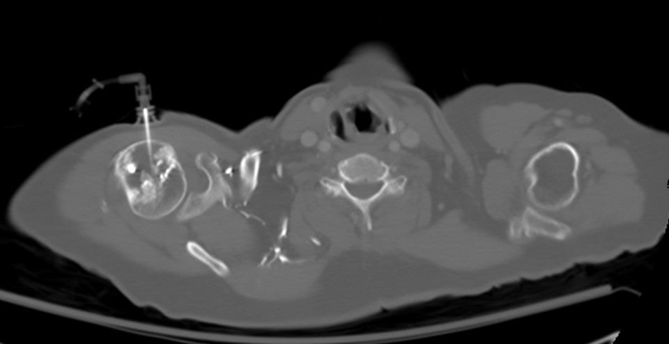
A needle in the top of an upper arm bone.
Scan: JasonRobertYoungMD, CC BY-SA 4.0
DRIVING QUESTION
What is a living long bone made of, and what does each part do?
START HERE
What Happened Here?
Finished observing? Click here to find out.
This is called intraosseous access. "Intra" means inside, and "osseous" means bone. Paramedics use it when a vein cannot be found fast enough, such as when a heart has stopped.
Today, you will find every part of a long bone. You will test why the shaft is a tube. Then you will push a model needle through the parts and see what it reaches.
BEFORE YOU START
What you will learn
I can label the parts of a long bone.
I can describe the function of each part.
I can trace a needle from the skin to the marrow and explain why it reaches the blood.
BEFORE YOU START
Get set up
1
Open mrsajdak.com/longbone on your device.
2
Write your name, the date and your period on the worksheet.
3
Find the three tabs at the top of the lab. Each tab goes with one part of your sheet.

| Tab in the lab | Worksheet | Questions |
|---|---|---|
| 1. Find the parts | Part 1 | 1 to 4 |
| 2. Test the shaft | Part 2 | 5 to 11 |
| 3. Trace the needle | Part 3 and Explain it | 12 to 16 |
Screenshot: mrsajdak.com/longbone
TAB 1 · QUESTIONS 1 TO 4
Part 1. Find the parts
A real femur, sawn the long way.
What is left on a cleaned bone? What did cleaning take away?
Photo: The American Journal of Anatomy (1917), public domain
PART 1 · QUESTIONS 1 AND 2
Visit every spot
1
Pick a letter, or click a yellow spot.
2
Look at it in the Zoomed in box.
3
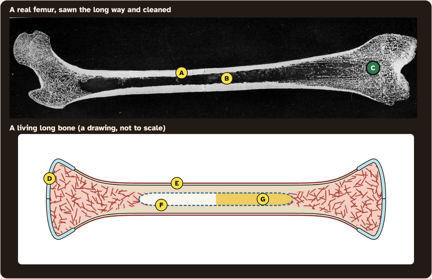
A to C are on the real bone. Tick what you see.
4
D to G are on the living bone. Tick what each one does.
PART 1 · LEARN IT · 1 OF 4
The two regions
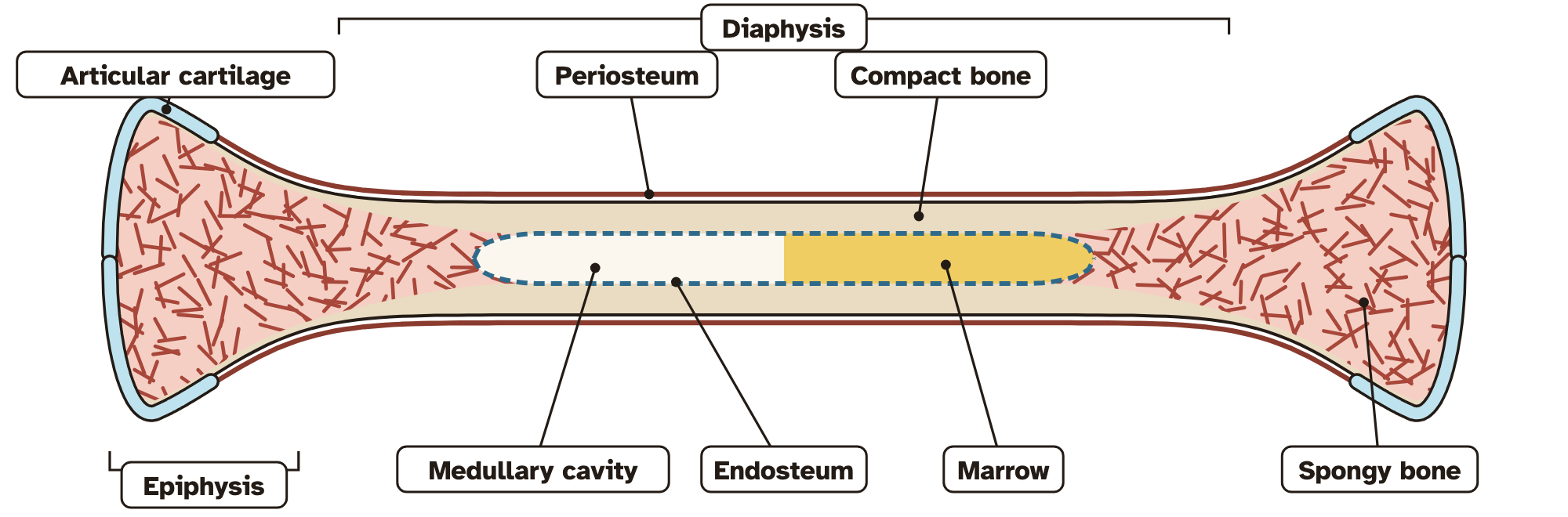
The long middle is the diaphysis, the shaft. Each wide end is an epiphysis. An epiphysis meets another bone at a joint.
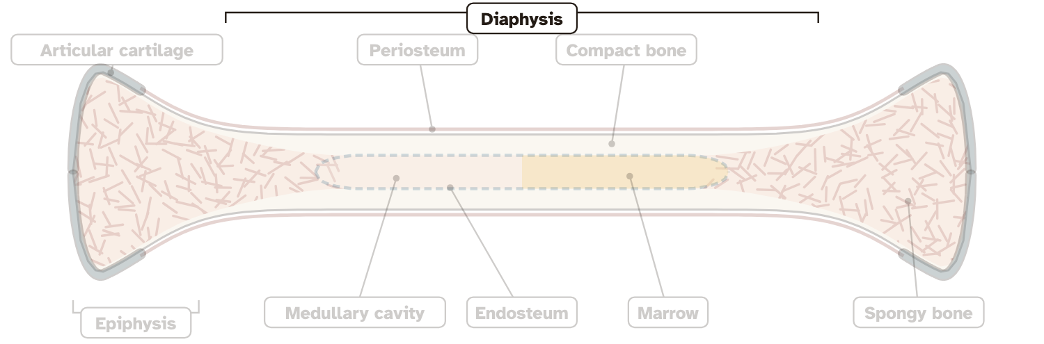
Drawing: Ryan Sajdak
PART 1 · LEARN IT · 2 OF 4
The two hard tissues
The solid wall of the shaft is compact bone. The net of thin plates inside each end is spongy bone. You saw both in spots A and C.
Photo: The American Journal of Anatomy (1917), public domain
PART 1 · LEARN IT · 3 OF 4
What a cleaned bone is missing
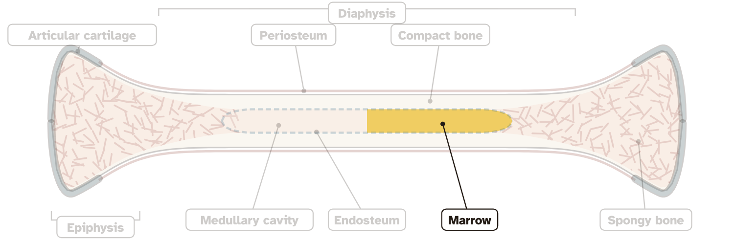
Articular cartilage caps each end. Periosteum wraps the outside. Endosteum lines the cavity. Marrow fills the cavity and the spaces in spongy bone.
Drawing: Ryan Sajdak
PART 1 · LEARN IT · 4 OF 4
The hollow is not empty
The channel in the shaft is the medullary cavity. It looks empty in a cleaned bone. In a living bone, marrow fills it.
Drawing: Ryan Sajdak
PART 1 · QUESTION 3
Label the bone
1
Pick a word in Step 2.
2
Click its number on the bottom picture.
3
If it is wrong, look at where the line points.
4
Copy all nine onto your sheet.
PART 1 · QUESTION 4
Model check
The real bone
Name two parts cleaning took away.
The drawing
It is a model. Name one thing it does not show well.
Screenshot: mrsajdak.com/longbone, Tab 1
TAB 2 · QUESTIONS 5 TO 11
Part 2. Test the shaft
Is a tube really better than a solid rod?
Then: what is the job of each part?
Photo: The American Journal of Anatomy (1917), public domain
PART 2 · QUESTIONS 5 AND 6
Measure the shaft
1
Pick line O. Click one edge, then the other. Press Check.
2
Do the same for line C.
3
Write the lab's numbers.
4
Wall = (outer − cavity) ÷ 2.
PART 2 · QUESTION 6
Find the wall
QUESTION 6
Why divide by 2?
The outer width crosses the wall two times, once on each side of the cavity.
Example
A pipe is 20 mm wide. Its hole is 8 mm wide.
(20 − 8) ÷ 2 = 6 mm
These are not your numbers. Use your own.
PART 2 · QUESTIONS 7 AND 8
Same bone, different tube
1
Set the slider to 0. Press Record this tube.
2
Do the same at 11, 20 and 30.
3
Copy your records.
4
What happens to the strength? What happens to the wall?
PART 2 · LEARN IT · 1 OF 4
Why the shaft is a tube
A tube puts the bone far from the middle, where bending pushes and pulls hardest. So the medullary cavity saves weight and makes room for marrow.
Photo: The American Journal of Anatomy (1917), public domain
PART 2 · LEARN IT · 2 OF 4
Jobs at the ends
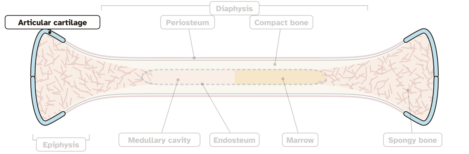
Epiphysis: the wide end meets another bone and forms a joint. Articular cartilage: a smooth, slippery cap that lets the joint move easily and cushions it.
Drawing: Ryan Sajdak
PART 2 · LEARN IT · 3 OF 4
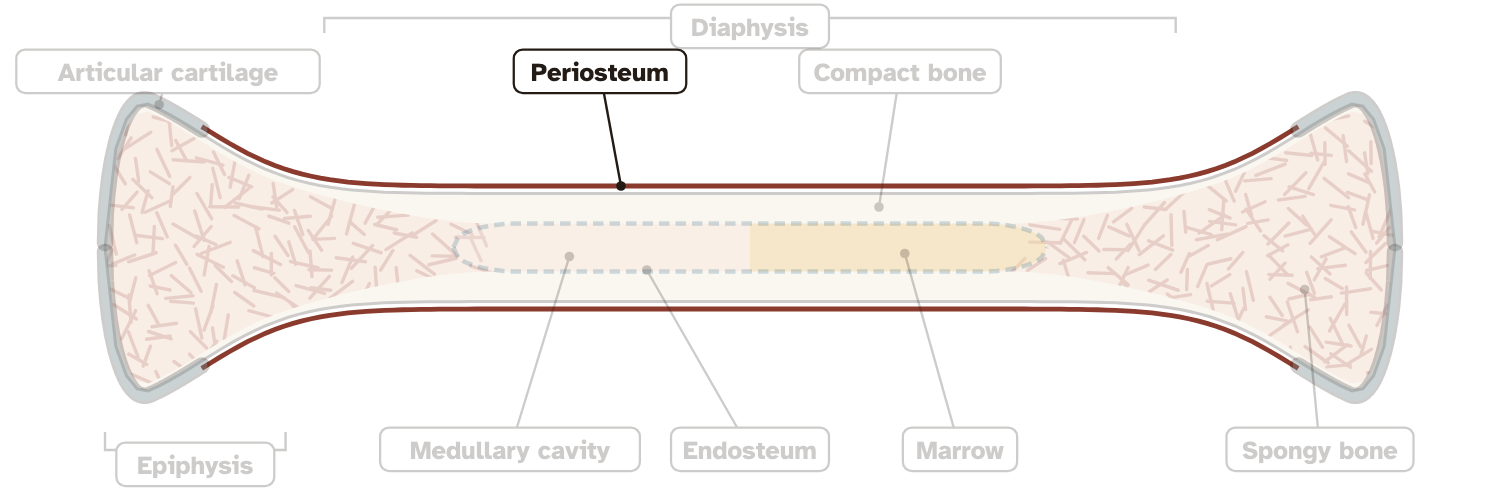
Jobs on the outside
Periosteum: a tough sleeve. Tendons and ligaments blend into it, and it helps form and repair bone. Compact bone: a strong wall that resists bending.
Drawing: Ryan Sajdak
PART 2 · LEARN IT · 4 OF 4
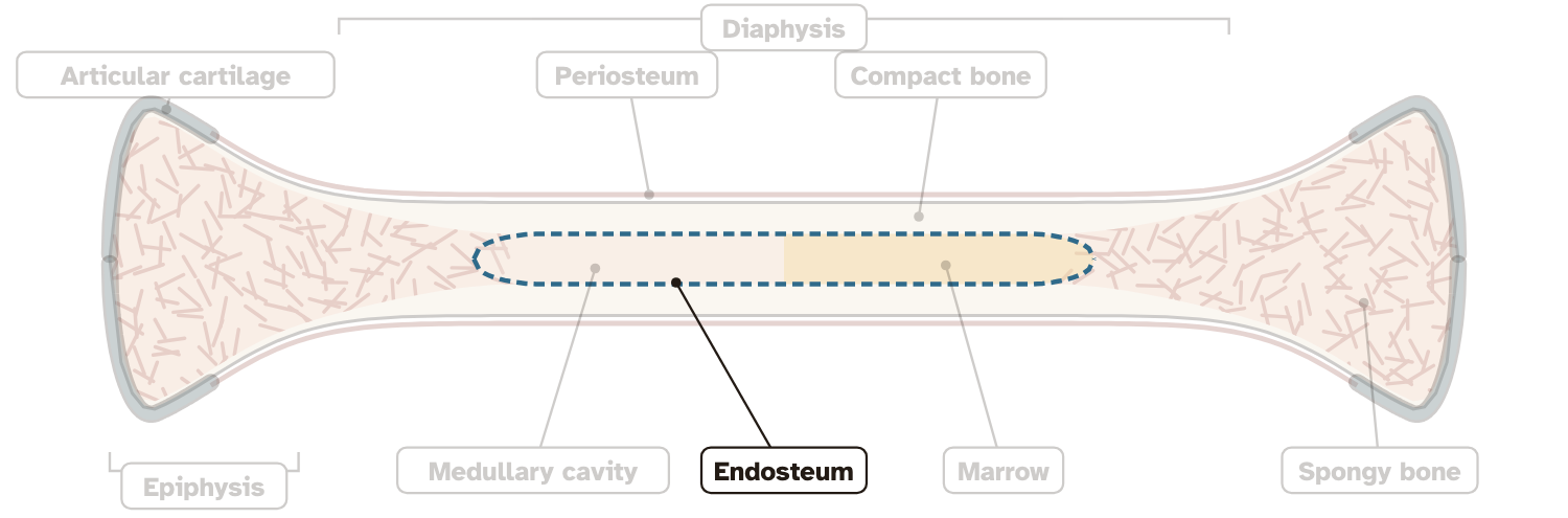
Jobs on the inside
Endosteum: a thin lining with bone-forming cells. Spongy bone: light, with spaces for red marrow. Red marrow makes blood cells. Yellow marrow stores fat.
Drawing: Ryan Sajdak
PART 2 · QUESTIONS 9 TO 11
Use the idea
QUESTION 9
Jobs
One job for each part
A job says what it does, not what it looks like.
QUESTION 10
Cases
Which part failed?
Do the four cases in the lab.
QUESTION 11
Limits
Model check
Why are real femurs not built like the 30 mm tube?
TAB 3 · QUESTIONS 12 TO 16
Part 3. Trace the needle
A shin bone is a long bone too.
Push a model needle in at two sites.
Photo: JasonRobertYoungMD, CC BY-SA 4.0
PART 3 · QUESTIONS 12 AND 13
Push the needle
1
Pick Near the end.
2
Move the slider slowly to 25 mm. Watch the push.
3
Pick Middle of the shaft. Do the same.
4
Thickness = drop depth − jump depth.
PART 3 · LEARN IT · 1 OF 3
What the needle went through
First skin and fat. Next the periosteum. Then compact bone, the hard part, which is why the needle has a drill. Then the push drops: the tip is in marrow.
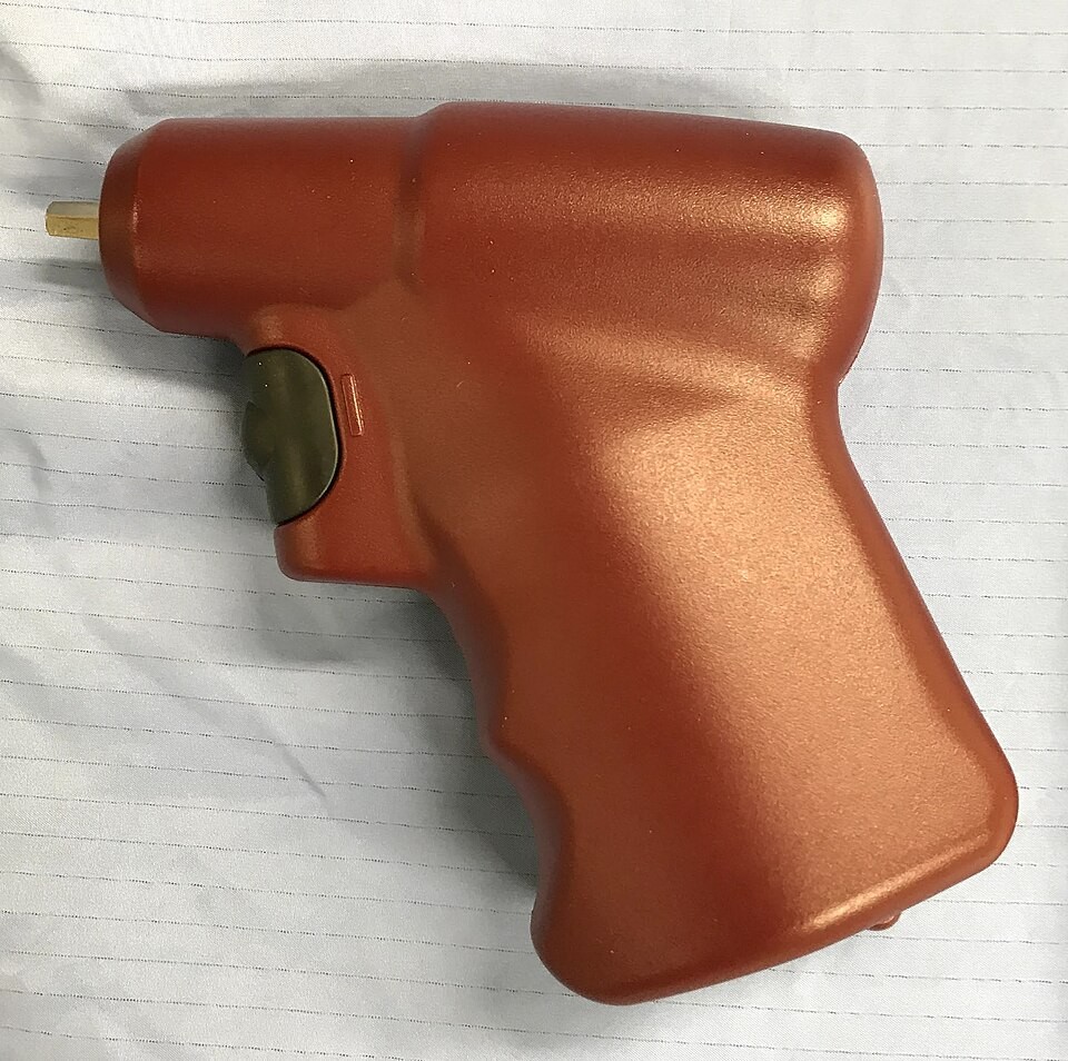
Photo: James Heilman MD, CC BY-SA 4.0
PART 3 · LEARN IT · 2 OF 3
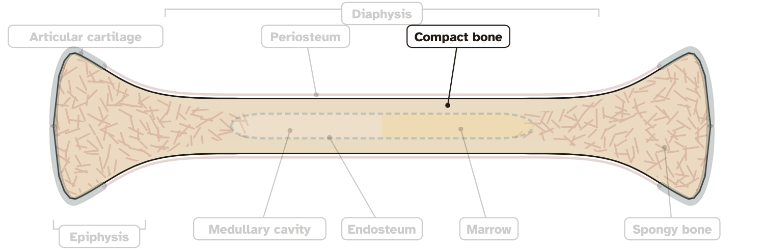
Why near the end?
Near the end, the wall of compact bone is thin. A thin wall is quicker to cross. Inside it, the needle sits in spongy bone full of marrow.
Drawing: Ryan Sajdak
PART 3 · LEARN IT · 3 OF 3
From marrow to the heart
Marrow is full of small blood vessels. They drain into veins that leave the bone. Medicine in the marrow goes the same way, into the veins and on to the heart.
Photo: JasonRobertYoungMD, CC BY-SA 4.0
EXPLAIN IT · QUESTION 16
Why does a needle in a bone reach the heart?
What I saw
Where the push changed, with a number.
The science idea
What is at the tip, and what it is full of.
So
How the medicine gets to the heart.
The tip of the needle sits inside the bone.
Scan: JasonRobertYoungMD, CC BY-SA 4.0
WRAP IT UP · BACK TO THE START
Revisiting the Phenomenon
What is a living long bone made of, and what does each part do?
1Using evidence from today, is a bone dry and dead? How do you know?
2What fills the hollow in a living shaft?
3Name three parts and the job of each.
4Which number from your worksheet best supports your answer?
Drawing: Ryan Sajdak
LAST CHECK
Before you hand it in
Name, date and period are on top.
All nine parts are labeled.
Every table is full.
Explain it has all three parts.
Photo: U.S. Navy, Seaman Justin Ontivero, public domain