ANATOMY & PHYSIOLOGY · SECTION 8.2
Inside a Muscle Fiber
Follow along with your worksheet, one question at a time.
mrsajdak.com/muscle-fiber
Photo: Peter Paplanus, CC BY 2.0
START HERE · PICK AN OPENER
Choose how to open the lesson
Click a card to jump to it. Every option ends at the same Observe, Wonder, Predict slide and the same driving question.
OPTION A · VIDEO
Faster Than a Blink
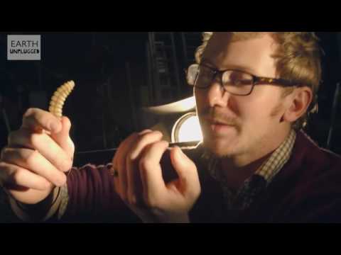
Clip 1 is a rattlesnake rattle filmed in slow motion (Earth Unplugged, a re-upload). Clip 2 is Deep Look on how the rattle works. Clip 3 is a Deep Look short of a hummingbird hovering.
Watch for
How fast do you think the tail muscle switches on and off?
Could your arm muscle do that? For how long?
What might be different inside a muscle cell that is this fast?
Videos on YouTube: YouTube re-upload; Deep Look.
OPTION B · TRY IT · 3 MINUTES
Tap Race
1
Pair up. One partner taps one finger on the desk as fast as they can.
2
The other partner counts the taps for 10 seconds.
3
Divide the taps by 10. That is your taps per second.
4
Switch jobs. Then try it for 30 seconds. Does your finger slow down?
You need
A desk and a partner
A clock or phone timer
Watch for
How many taps a second did you get?
A rattlesnake gets about 90. How many times faster is that?
Did your finger get tired? What might a muscle cell need so it never tires?
OPTION C · PHOTOS
Phenomenon: Same Parts, Different Jobs
Photos: Peter Paplanus, CC BY 2.0 · LexSprinter, CC BY-SA 3.0
START HERE
Observe, wonder, predict
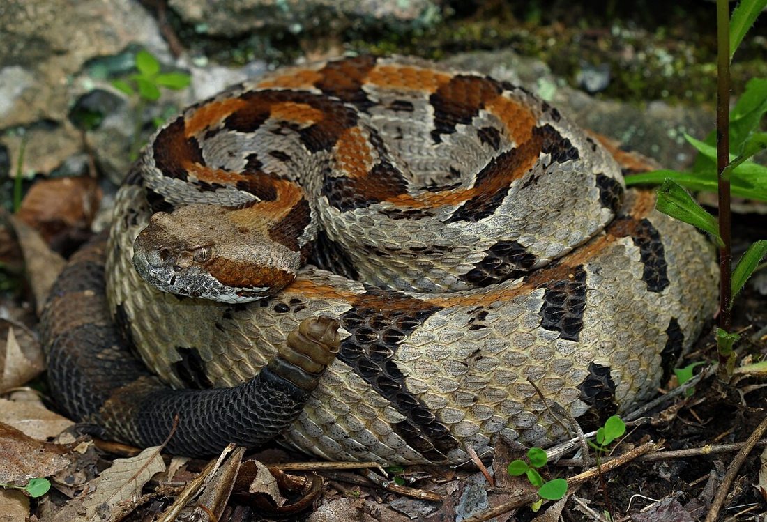
A rattlesnake's tail muscle shakes about 90 times a second and does not tire. A sprinter's thigh pushes very hard, but only for a few seconds.
OBSERVEWhat does each muscle do well?
WONDERWhat do you wonder about the inside of a muscle cell?
PREDICTAre the two muscle cells built the same way inside? Why or why not?
Photo: Peter Paplanus, CC BY 2.0
DRIVING QUESTION
How can one muscle cell be built to shake a rattle 90 times a second, while another lifts your whole body?
START HERE
What Happened Here?
Finished observing? Click here to find out.
Every skeletal muscle cell has the same parts. But the amount of each part is different, and that changes what the muscle is good at.
Today, you will find every part of one muscle fiber. You will read the stripes in a real electron microscope picture. Then you will compare real muscles.
TAB 1 · QUESTIONS 1 TO 7
Part 1. Parts of a fiber
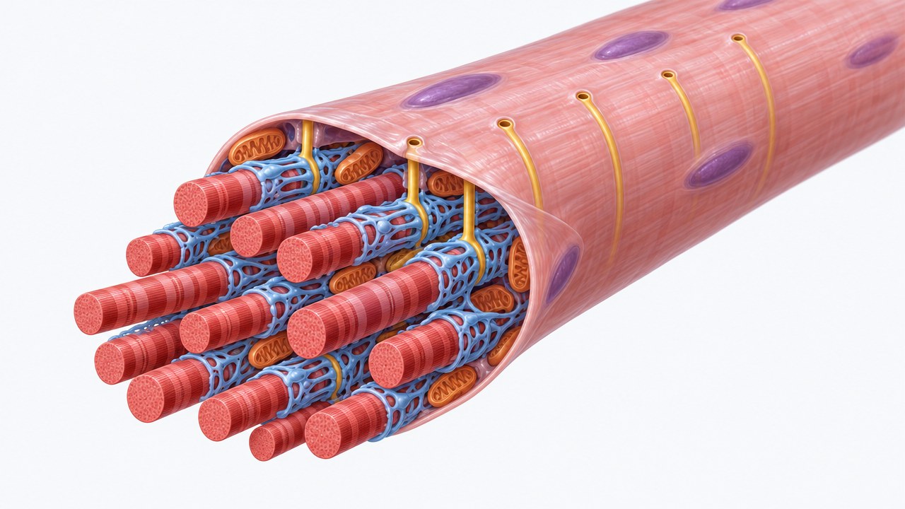
One muscle fiber is one long cell. What is inside it?
Photo: made for this lab (AI illustration)
PART 1 · QUESTIONS 1 TO 4
Click every part
1
Click all six parts.
2
Write what each one looks like.
3
Do Step 2, Look closely.
4
Answer Questions 1 to 4.
PART 1 · LEARN IT · 1 OF 3
The outside and the tubes
Sarcolemma: the cell membrane of a muscle fiber. A nerve signal spreads along it.
T-tubule (transverse tubule): a tube of sarcolemma that dips deep into the fiber. It carries the signal inside, so every part gets it at once.
Sarcoplasmic reticulum (SR): a net of sacs around each myofibril. It stores calcium and lets it out when the signal arrives.
Drawing: made for this lab (AI illustration)
PART 1 · LEARN IT · 2 OF 3
The inside
Myofibril: a long striped rod. Myofibrils fill most of the fiber. They are the parts that pull.
Mitochondrion: makes ATP, the energy the myofibrils use to pull.
Nucleus: holds the DNA. A fiber is so long that it has many nuclei, pressed to the edge.
Sarcoplasm: the cytoplasm of a muscle fiber. All of these parts sit in it.
Drawing: made for this lab (AI illustration)
PART 1 · LEARN IT · 3 OF 3
Every part has a job
The signal comes in on the sarcolemma and goes deep inside on the T-tubules. The SR holds calcium right next to every myofibril. The myofibrils pull. The mitochondria feed them energy. Structure fits function: each part's shape matches its job.
Drawing: made for this lab (AI illustration)
PART 1 · QUESTIONS 5 TO 7
Use the idea
QUESTION 5
Which part does each job?
Tick one part for each job.
Then do Match the job in the lab.
QUESTION 6
Label the drawing
Write the name of each lettered part, A to F.
QUESTION 7
Model check
Why does a long fiber need T-tubules, and not just the sarcolemma?
TAB 2 · QUESTIONS 8 TO 13
Part 2. Read the stripes
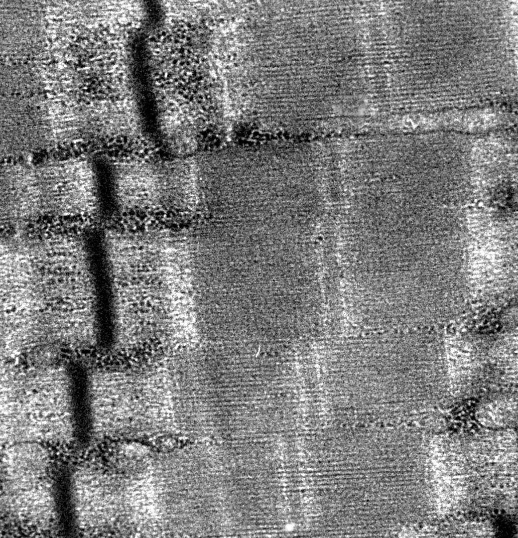
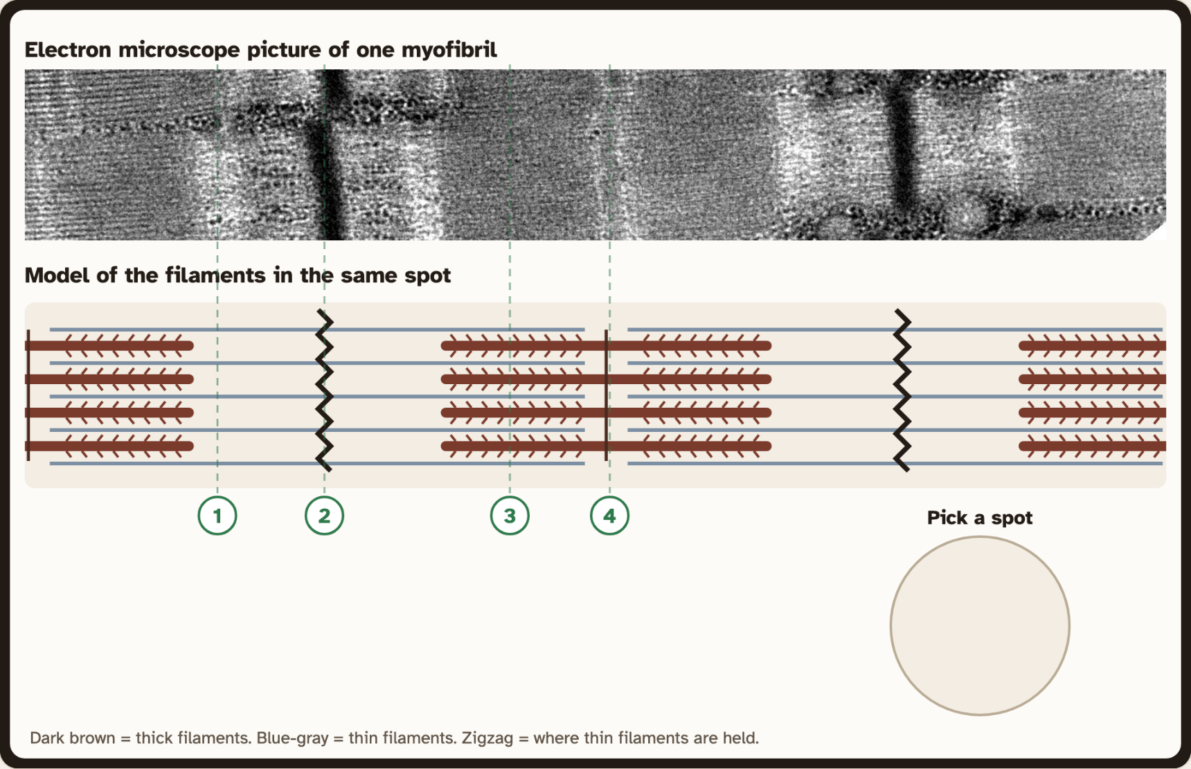
A real electron microscope picture of myofibrils. Why is it striped?
Photo: Louisa Howard, public domain
PART 2 · QUESTIONS 8 TO 10
Check each spot
1
Click spots 1 to 4.
2
Copy the darkness and the filaments.
3
Find the darkest spot.
4
Answer Questions 8 to 10.
PART 2 · QUESTION 10
What makes a spot dark?
THE IDEA
Stack it up
An electron beam shines through a thin slice.
Where more protein is in the way, less gets through. That spot looks darker.
Example
One sheet of paper on a window lets light through.
Three sheets let less through.
More layers = darker
These are not your numbers. Use your own.
PART 2 · LEARN IT · 1 OF 3
Two kinds of filaments
Thick filament: made of the protein myosin.
Thin filament: made mostly of the protein actin.
Sarcomere: one unit of a myofibril, from one Z line to the next. A myofibril is thousands of sarcomeres in a row.
Micrograph: Louisa Howard, public domain, with a model drawn for this lab
PART 2 · LEARN IT · 2 OF 3
The bands
Z line: the darkest line. It holds the thin filaments in place.
I band: the light band. Only thin filaments are there.
A band: the dark band. It is as long as the thick filaments.
H zone: the lighter middle of the A band. Only thick filaments are there.
M line: the line in the middle that holds the thick filaments in place.
Micrograph: Louisa Howard, public domain, with a model drawn for this lab
PART 2 · LEARN IT · 3 OF 3
Why the stripes look the way they do
More protein in the way makes the picture darker. The A band is dark because the thick filaments are there. Where thin filaments overlap them, it is darker still. The I band is light because it has only thin filaments. How these filaments work together is the next lesson.

Micrograph: Louisa Howard, public domain
PART 2 · QUESTIONS 11 TO 13
Name what you measured
QUESTION 11
Name each spot
I band, Z line, A band or H zone.
Use your records: which filaments are there?
QUESTION 12
Label the sarcomere
Write a name in each box.
QUESTION 13
Fill the blanks
Which protein is in each filament?
Where does one sarcomere start and stop?

TAB 3 · QUESTIONS 14 TO 18
Part 3. Built for the job
Real muscles, real measurements. Same parts, different mix.
Photo: Pslawinski, CC BY-SA 4.0
PART 3 · QUESTIONS 14 TO 16
Compare five muscles
1
Click all three parts.
2
Copy the records table.
3
Do the math in Step 2.
4
Answer Questions 14 to 16.
PART 3 · QUESTION 15
How many times as much?
THE MATH
Divide
Big amount ÷ small amount.
That is how many times as much.
Example
A jar holds 12 cups. A mug holds 3 cups.
12 ÷ 3
= 4 times as much
These are not your numbers. Use your own.
PART 3 · LEARN IT · 1 OF 2
Structure fits function
Every skeletal muscle fiber has the same parts. The mix is different. Lots of myofibrils: the fiber can pull hard. The human thigh is 85% myofibrils. Lots of mitochondria: the fiber can work for a long time without tiring. A hovering hummingbird needs that. Lots of SR: calcium goes out and back fast, so the fiber can switch on and off many times a second, like a rattle.
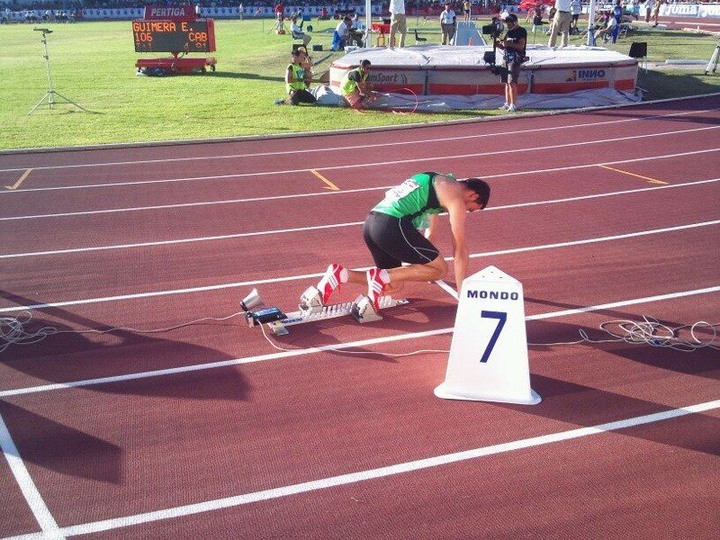
Photo: LexSprinter, CC BY-SA 3.0
PART 3 · LEARN IT · 2 OF 2
There is only so much room
A fiber is full. If one part takes more room, another gets less. The rattlesnake tail is about one third myofibrils. The human thigh is 85%. So the rattle muscle is fast and does not tire, but it is weak. That is a trade-off.
Photo: Peter Paplanus, CC BY 2.0
PART 3 · QUESTION 17
The trade-off
QUESTION 17
Why is the rattle muscle weak?
Use the numbers in your records table.
What takes up the room in the fiber?
IN THE LAB
What would it need?
Do the four cases under the Learn It box.
EXPLAIN IT · QUESTION 18
Marathon runner or weightlifter?
Claim
Which needs more mitochondria? Which needs more myofibrils?
Evidence
Use two numbers from the lab.
Reasoning
Say what each part does, and why there is a trade-off.
WRAP IT UP · BACK TO THE START
Revisiting the Phenomenon
How can one muscle cell be built to shake a rattle 90 times a second, while another lifts your whole body?
EVIDENCEWhich part lets the rattle switch on and off so fast?
EVIDENCEWhich part lets it shake for hours?
EVIDENCEWhat does it give up?
Hand in your worksheet.
Next lesson: what happens, step by step, when a fiber gets the signal.