ANATOMY & PHYSIOLOGY · SECTION 7.4
Two Ways to Build a Bone
Follow along with your worksheet, one question at a time.
mrsajdak.com/ossification
X-ray: Jason Hickey, CC BY 3.0
START HERE · PICK AN OPENER
Choose how to open the lesson
Click a card to jump to it. Every option ends at the same Observe, Wonder, Predict slide and the same driving question.
OPTION A · VIDEO
A Child's Broken Bone
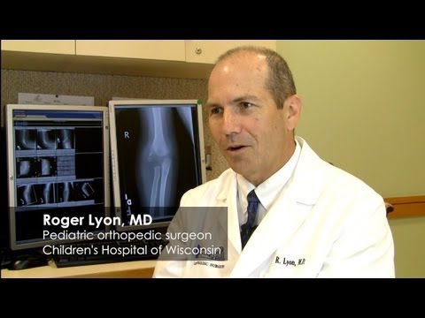
Clip 1 is a children's hospital surgeon on why a child's broken bone needs special care. Clip 2 is a short look at how a bone grows.
Watch for
What part of a child's bone worries the doctor?
Why would an adult not have this worry?
What do you think that part of the bone is made of?
Videos on YouTube: Children's Wisconsin; Children's Hospital Colorado.
OPTION B · TRY IT · 3 MINUTES
The Cartilage You Still Have
1
Press the bridge of your nose, up high. Try to bend it. Do not force it.
2
Now press the soft tip of your nose. Bend it gently.
3
Bend the top of your ear gently. Let go.
4
Which places bend and spring back? Which places do not move?
You need
Your own nose and ear
Nothing else
Watch for
The bridge of the nose is hard and does not bend.
The tip of the nose and the ear bend and spring back.
What would it be like if your whole skeleton still felt like that?
OPTION C · PHOTOS
Phenomenon: Two Gaps, No Breaks
Photos: Johann Dréo, CC BY-SA 3.0 · Jason Hickey, CC BY 3.0
START HERE
Observe, wonder, predict
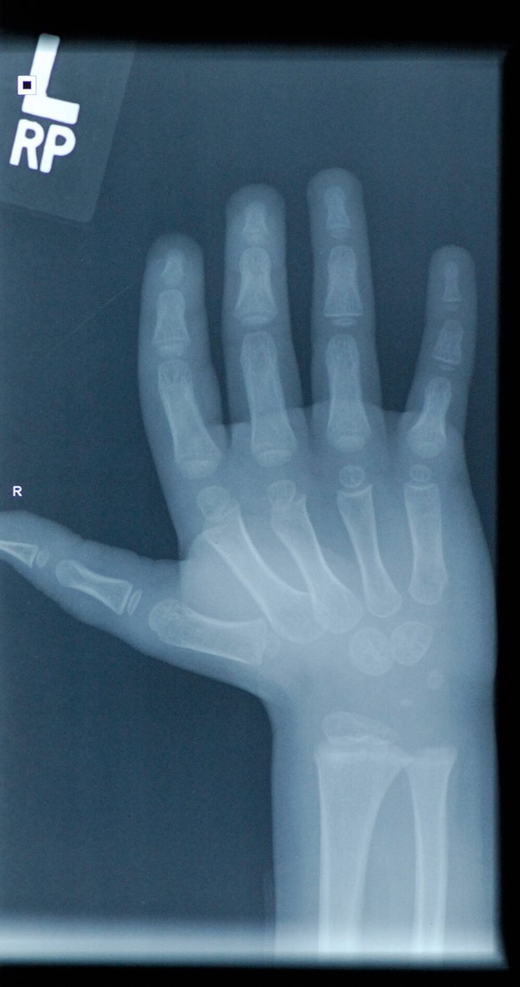
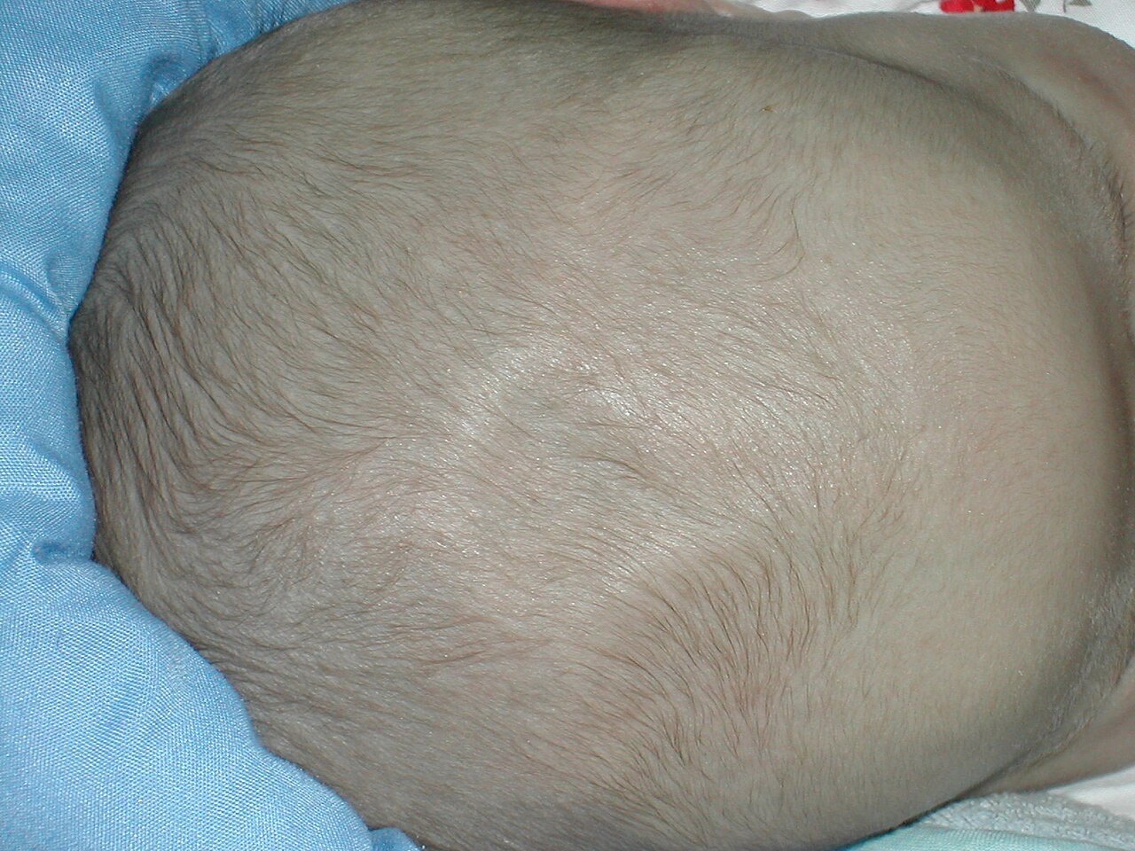
A newborn's skull has a soft spot with no bone in it. A young X-ray shows dark gaps near the ends of the bones.
OBSERVEWhere exactly is each gap?
WONDERWhat do you wonder about how bones form?
PREDICTWhat fills each gap?
Look where the end of a bone meets its shaft.
X-ray: Jason Hickey, CC BY 3.0
DRIVING QUESTION
How can a newborn's skull and a teen's leg bone both be unfinished?
START HERE
What Happened Here?
Finished observing? Click here to find out.
Both gaps are normal. A skeleton is built over many years, and neither of these bones is done yet.
Today, you will watch two bones form. You will test how a bone gets longer against a real experiment from 1727. Then you will read real X-rays.
TAB 1 · QUESTIONS 1 TO 6
Part 1. Watch two bones form
A skull bone and a leg bone. What is there first?
Photo: Johann Dréo, CC BY-SA 3.0
PART 1 · QUESTIONS 1 TO 3
Click every stage
1
Click stages 1 to 4 of the skull bone.
2
Click stages 1 to 6 of the leg bone.
3
Read what each stage says.
4
Answer Questions 1 to 3.
PART 1 · LEARN IT · 1 OF 5
Ossification
Ossification is the forming of bone. It happens in two ways.
Drawing: Ryan Sajdak
PART 1 · LEARN IT · 2 OF 5
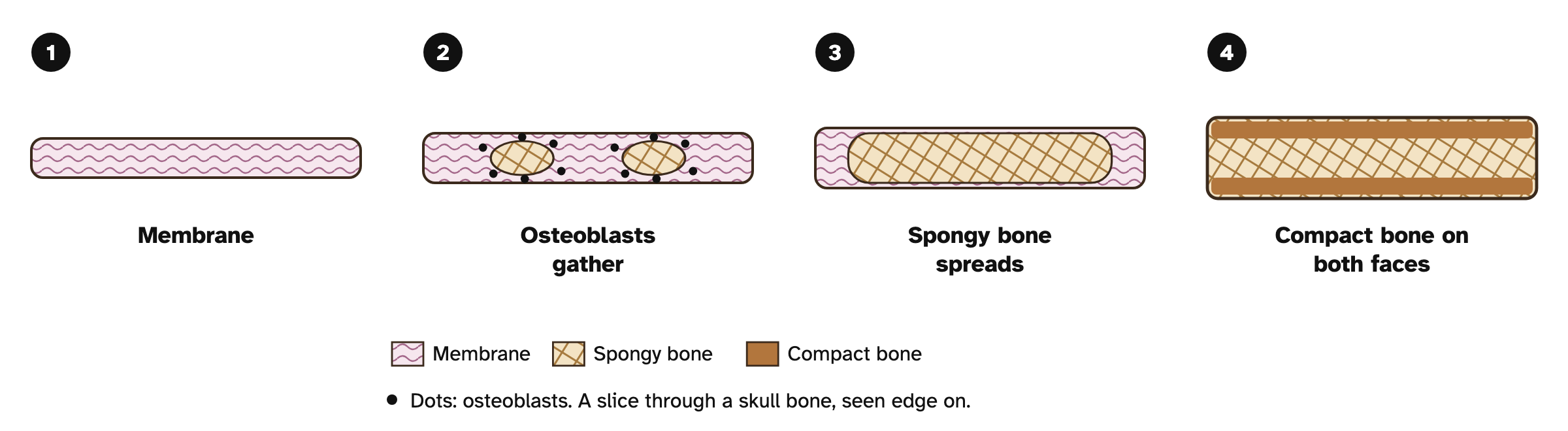
Intramembranous
The skull bone started as a sheet of membrane. Osteoblasts lay down bone right there. There is no cartilage.
Drawing: Ryan Sajdak
PART 1 · LEARN IT · 3 OF 5
Endochondral
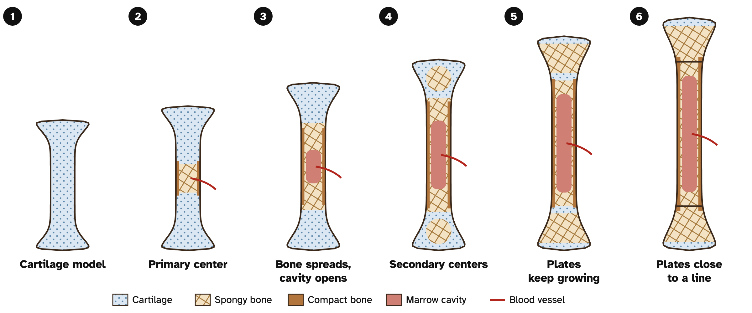
The leg bone started as a cartilage model. Bone forms first at the primary ossification center, then at secondary ossification centers.
Drawing: Ryan Sajdak
PART 1 · LEARN IT · 4 OF 5
The centers on a real X-ray
The bright shaft is the primary center. The small bright piece in the end is a secondary center. The dark band is cartilage.
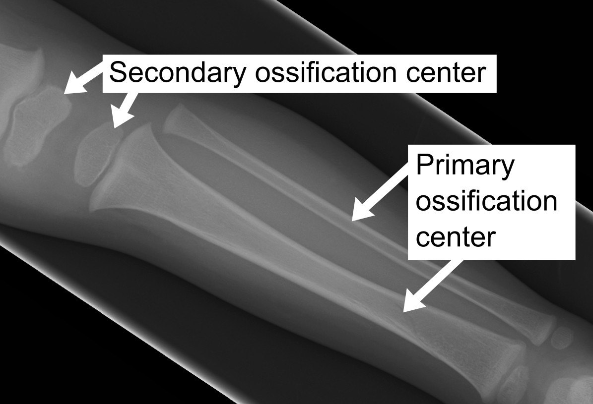
Photo: Mikael Häggström, from a source image by James Heilman, CC BY-SA 3.0
PART 1 · LEARN IT · 5 OF 5
Cartilage does not turn into bone
The cartilage breaks down. Osteoclasts clear what is left. Osteoblasts build new bone in its place.
Drawing: Ryan Sajdak
PART 1 · QUESTIONS 4 TO 6
Order, sort, compare
1
Click the cards in order, youngest first.
2
Sort the bones.
3
Fill in the compare table.
TAB 2 · QUESTIONS 7 TO 11
Part 2. How does it get longer?
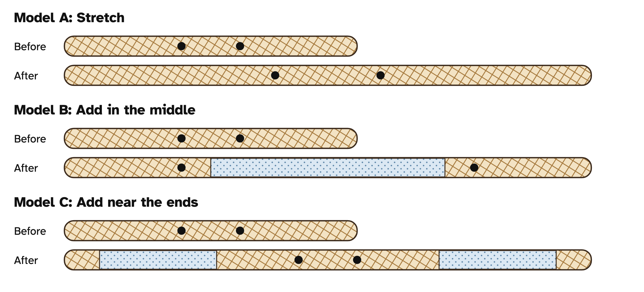
Three ideas. Two marks on the shaft.
Photo: Jason Hickey, CC BY 3.0
PART 2 · QUESTIONS 7 AND 8
Test all three ideas
1
Predict in Question 7.
2
Pick an idea. Slide to week 8.
3
Press Record.
4
Do all three. Copy your records.
PART 2 · QUESTION 9
The real test, 1727
THE TEST
Stephen Hales
Two holes in the shaft of a growing chicken's leg bone.
He looked again two months later.
THE DATA
Now compare
The 1727 box opens in the lab after all three records.
Which idea matches his numbers?
PART 2 · LEARN IT · 1 OF 3
The marks did not move
The shaft did not stretch. New length was added near the ends, at the epiphyseal plates.
Drawing: Ryan Sajdak
PART 2 · LEARN IT · 2 OF 3
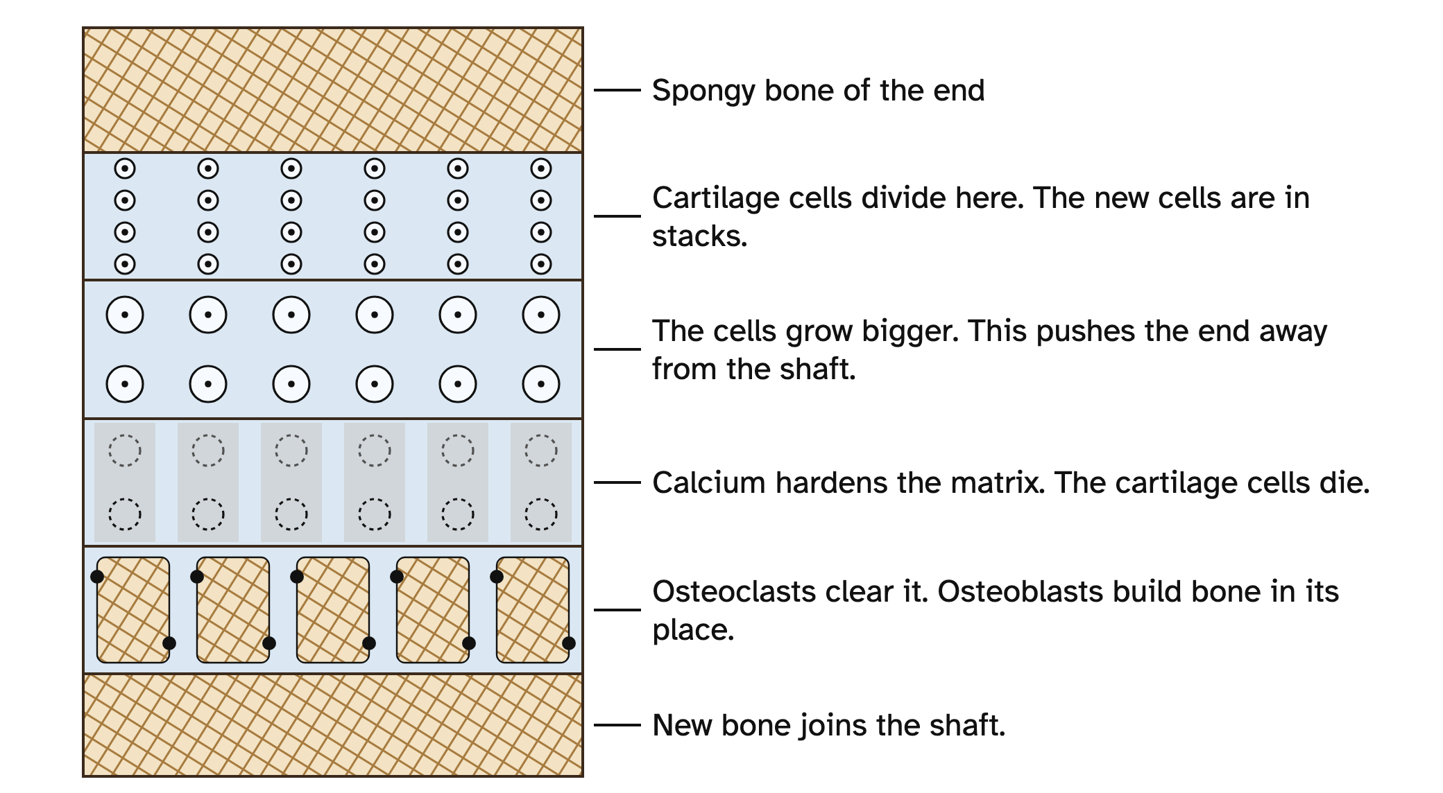
Inside the plate
Cartilage cells divide and grow bigger. On the shaft side, osteoclasts clear the hard cartilage and osteoblasts build bone.
Drawing: Ryan Sajdak
PART 2 · LEARN IT · 3 OF 3
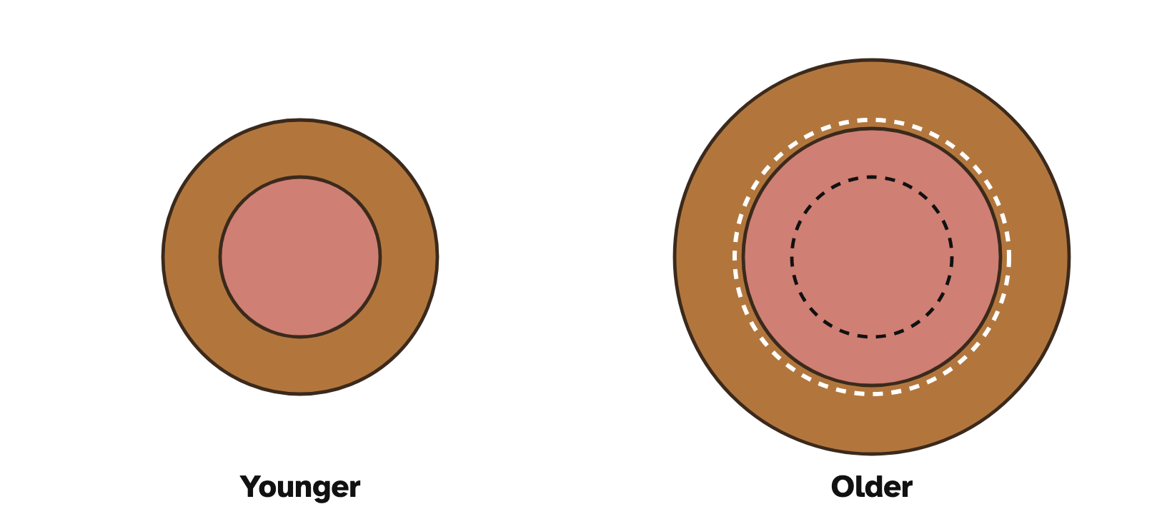
Getting wider
Osteoblasts under the periosteum add bone outside. Osteoclasts remove bone inside.
Drawing: Ryan Sajdak
PART 2 · QUESTIONS 10 AND 11
Use the idea
QUESTION 10
Number the steps
Go from the end of the bone toward the shaft.
QUESTION 11
Model check
SEP 2. Tab 2 is a model. Name one thing it leaves out.
TAB 3 · QUESTIONS 12 TO 16
Part 3. Read the plate
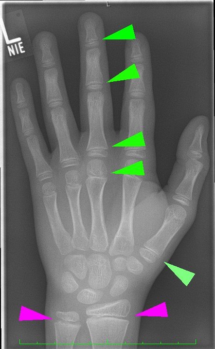
Real X-rays. Bone looks bright.
Photo: eigen, CC BY-SA 4.0
PART 3 · QUESTIONS 12 AND 13
Look at four X-rays
1
Pick an X-ray.
2
Look where the end meets the shaft.
3
Press Dark gap or Solid bone.
4
Answer Questions 12 and 13.
PART 3 · LEARN IT · 1 OF 2
Why the plate looks dark
Cartilage barely shows on an X-ray. So an open epiphyseal plate looks like a dark gap. It is not a break.
Photo: eigen, CC BY-SA 4.0
PART 3 · LEARN IT · 2 OF 2
When the plate closes
Late in the teens, bone replaces the plate. A thin epiphyseal line may remain. The bone can no longer get longer.
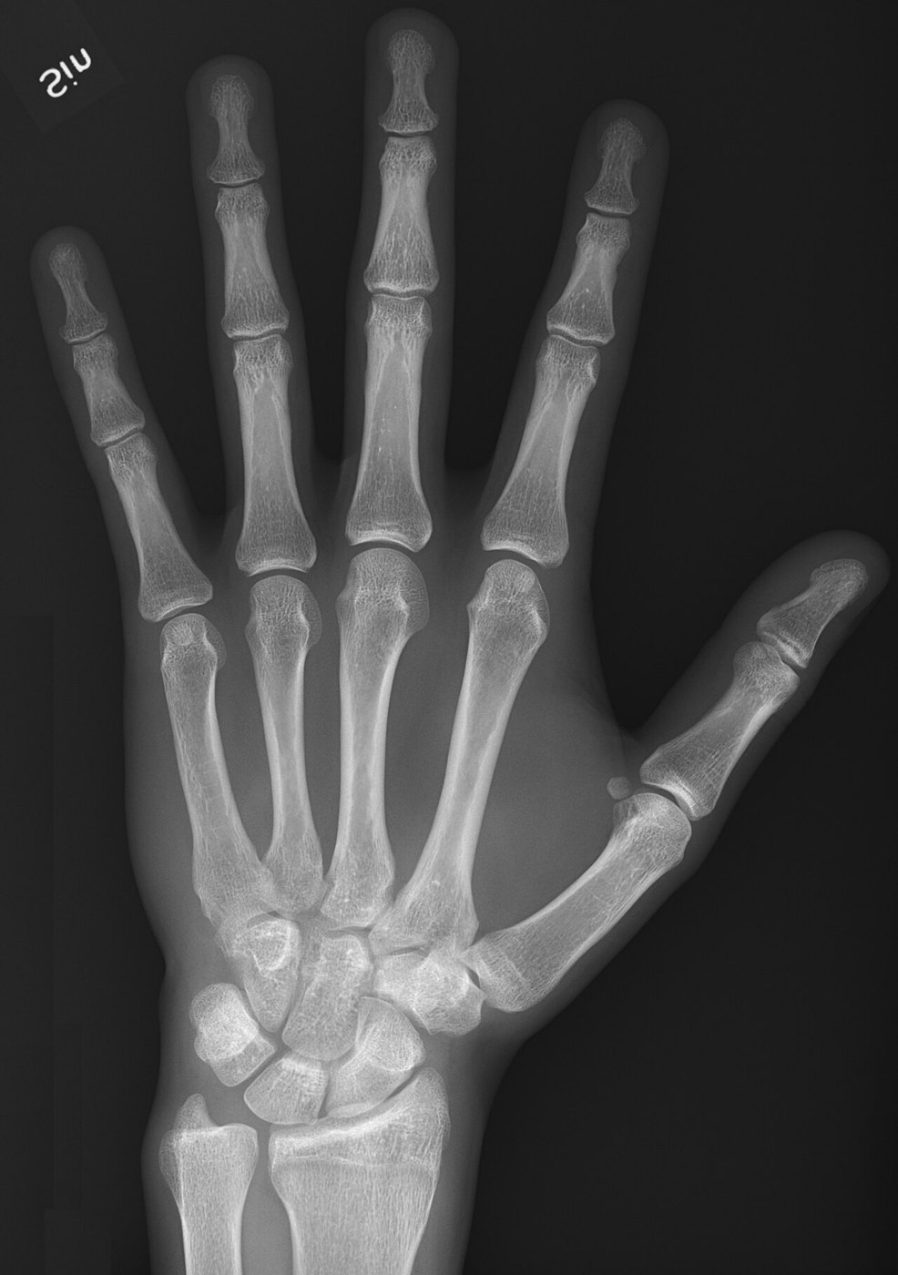
Photo: Mikael Häggström, CC0
PART 3 · QUESTION 14
The growth rule
THE RULE
16 mm a year
The two knee plates add about 10 mm + 6 mm a year.
Growth left = years until the plates close × 16 mm.
It fits only the last years of growth.
Example
Bone age 10. Plates close at 16.
16 − 10 = 6 years.
6 × 16 mm = 96 mm
These are not your numbers. Use your own.
EXPLAIN IT · QUESTION 16
How are the two gaps different?
What I saw
A soft spot on a skull, a dark gap near the end of a bone.
Science idea
What each gap is made of, and how each bone is built.
Explains it
Say how that makes the two gaps different.
WRAP IT UP · BACK TO THE START
Revisiting the Phenomenon
How can a newborn's skull and a teen's leg bone both be unfinished?
EVIDENCEWhich Tab 1 stage is the soft spot closest to?
EVIDENCEWhat did Hales's marks show?
EVIDENCEWhat does a dark gap tell you?
Hand in your worksheet.
Next lesson: synovial joints.