ANATOMY AND PHYSIOLOGY · CHAPTER 6
Skin Under the Microscope
Follow along with your worksheet, one question at a time.
mrsajdak.com/skin-layers
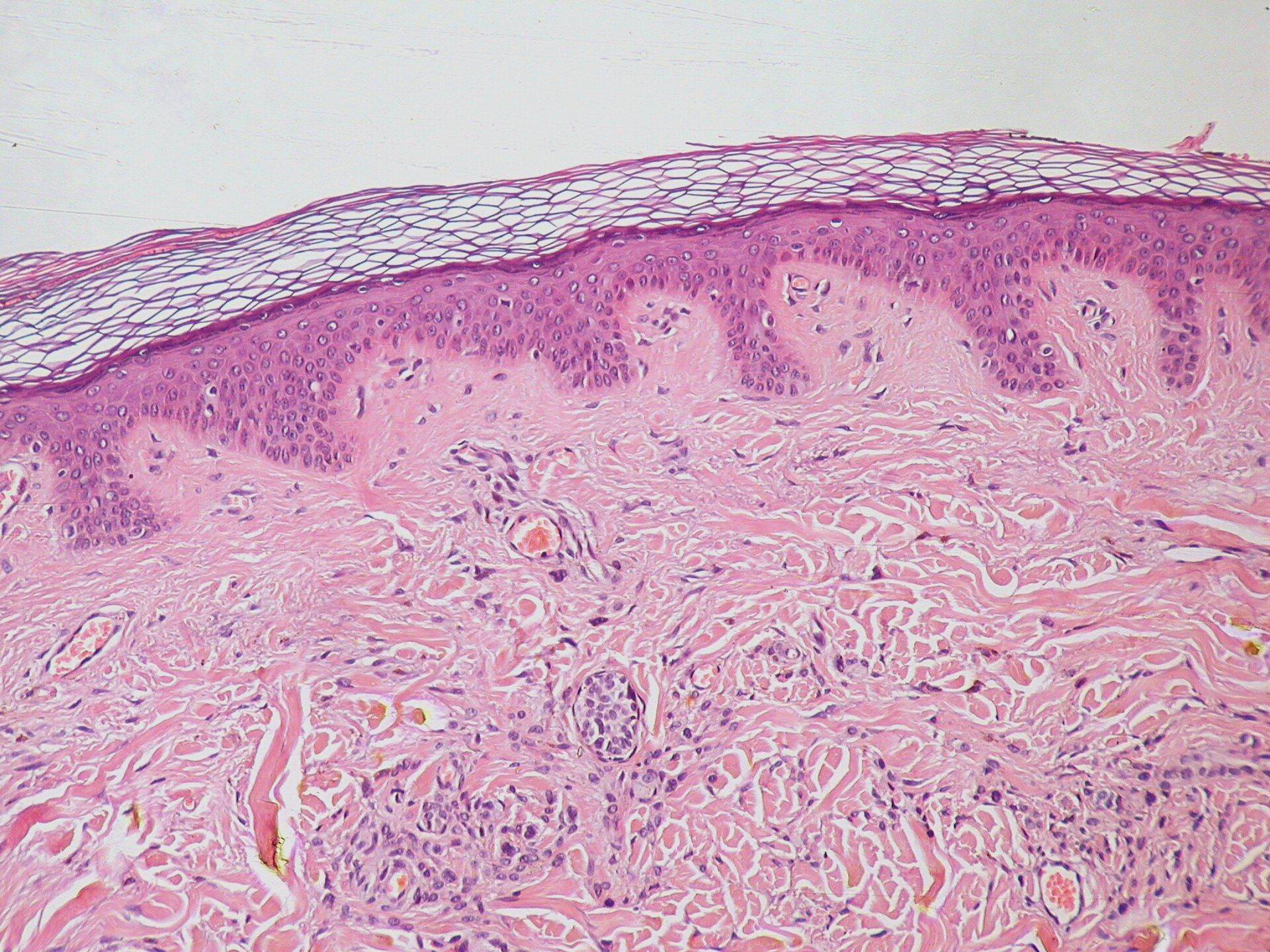
Photo: Kilbad, public domain
START HERE
Phenomenon: The Mark That Stays
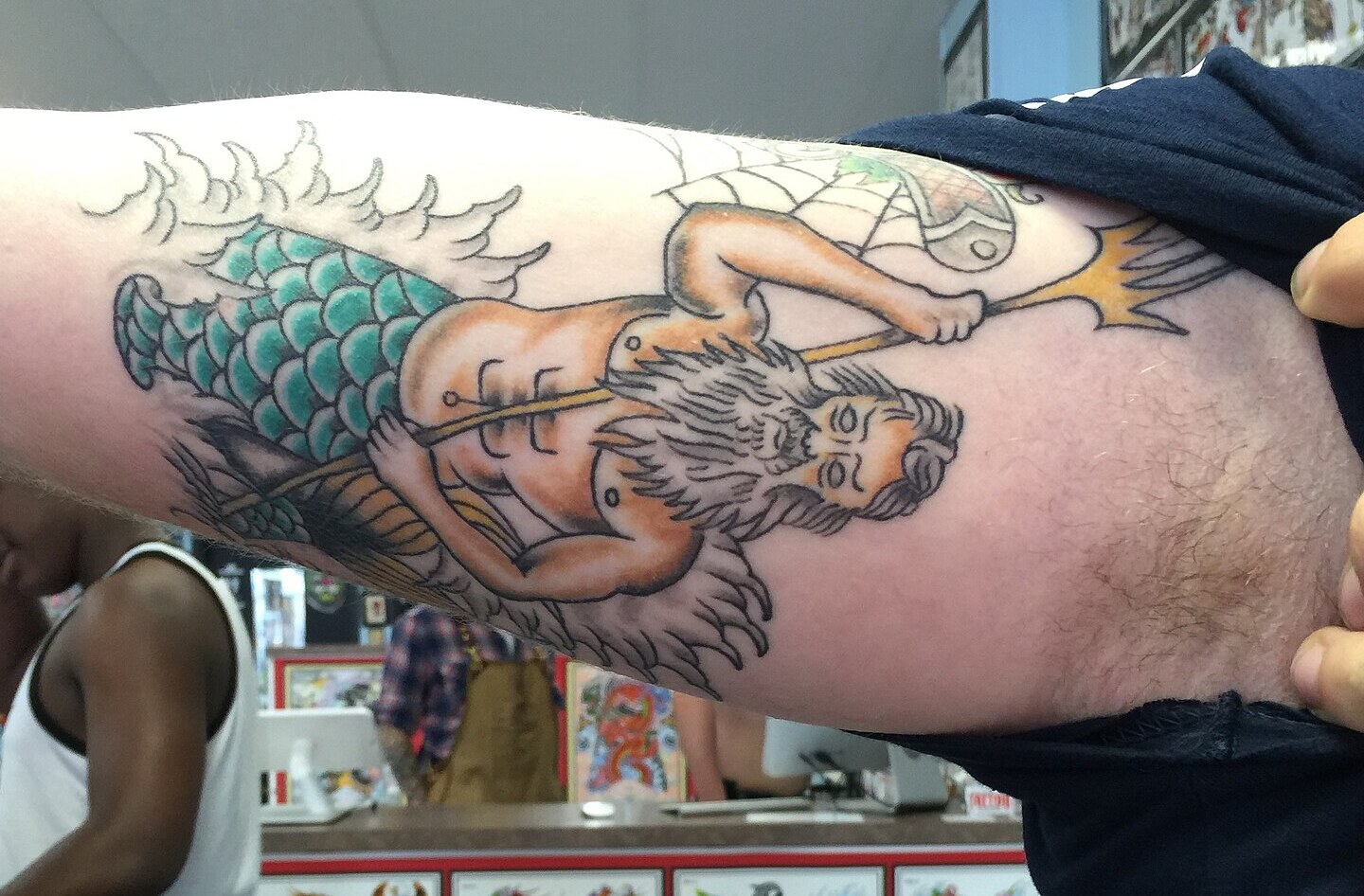
Photos: Tony Alter, CC BY 2.0
START HERE
Observe, wonder, predict
These photographs show two tattoos of the same sea god. One has sharp lines and bright color. The other is faded and blurred. Tattoos change slowly over many years, but they do not go away. Skin gets washed, rubbed and scraped every day. So why is the ink still there?
OBSERVEWhat differences do you notice between the two tattoos?
WONDERWhere in the skin might the ink be?
PREDICTWould a tattoo last if the ink went only into the very top of the skin? Explain your reasoning.
DRIVING QUESTION
Why does a tattoo stay when skin wears away?
START HERE
What Is Going On Here?
Finished observing? Click here to find out.
Your skin is always wearing away. Every minute, you lose tens of thousands of tiny skin cells. In about a month, the whole outer surface of your skin is replaced with new cells. Yet a tattoo can last a lifetime. It may fade and blur, but it does not go away.
Today, you will investigate what skin is made of. You will look at skin under a virtual microscope, measure how thick it is, compare six places on the body, and follow one skin cell through its whole life. At the end of the investigation, you will use your evidence to explain why a tattoo lasts and return to the original driving question.
BEFORE YOU START
Skin wears away. A tattoo stays.
The layers
1. Epidermis, on top
2. Dermis, below it
3. Subcutaneous layer, beneath the skin
Skin is the first two. Each layer is built for a different job.
The answer to the driving question is somewhere in these layers. Your job is to find it.
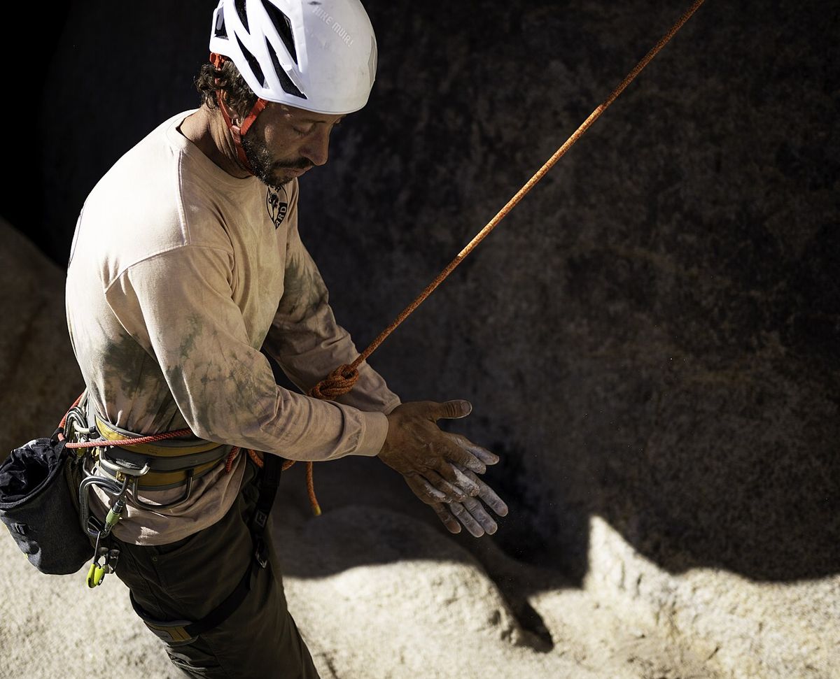
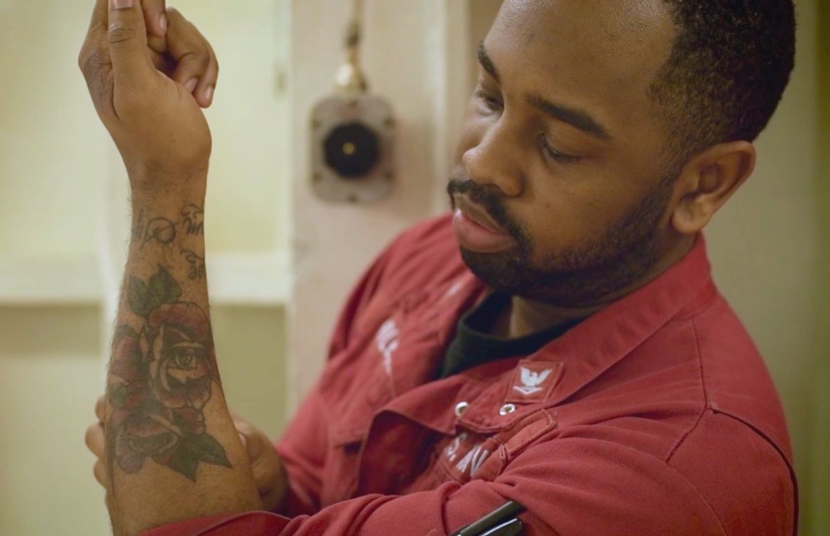
Photos: Emily Hassell, National Park Service · Brandon Richardson, U.S. Navy
BEFORE YOU START
What you will learn
DAY 1
I can identify the epidermis, dermis and subcutaneous layer on a slide and describe the role of each.
I can measure skin layers with a microscope scale and explain why skin is thicker in some places.
DAY 2
I can identify the five strata of the epidermis and what happens to a cell in each.
I can tell the papillary from the reticular layer and explain how each one's build fits its job.
BEFORE YOU START
Get set up
1
Open mrsajdak.com/skin-layers on your device.
2
Write your name, the date and your period on the worksheet.
3
Find the four tabs at the top of the lab. Each tab goes with one part of the worksheet.
4
Day 1 is the front of the worksheet. Day 2 is the back.
5
Start on Tab 1. Do not skip ahead.
| Tab in the lab | Worksheet | Questions |
|---|---|---|
| 1. Virtual microscope | Day 1 · Part 1 | 1 to 6 |
| 2. Body map | Day 1 · Part 2 | 7 to 10 |
| 3. Inside the epidermis | Day 2 · Part 3 | 11 to 15 |
| 4. Inside the dermis | Day 2 · Part 4, Wrap it up | 16 to 20 |
Screenshot: mrsajdak.com/skin-layers
DAY 1 · TAB 1 · QUESTIONS 1 TO 6
Part 1. Virtual microscope
A thin slice of skin sits on a slide. Stain makes its parts show up.
You will find the three layers. Then you will measure them and test three injuries.
Photo: Hennepin County Library, public domain
PART 1 · QUESTION 1
Focus, then find the layers
1
Pick a slide. Start with Thin skin · forearm.
2
Move the Focus slider until the lab says sharp.
3
Click a spot inside the circle. A yellow marker shows up.
4
Name the layer under the marker. A wrong pick gets crossed out.
5
Find all three layers on both slides. Then tick the clues in Question 1.
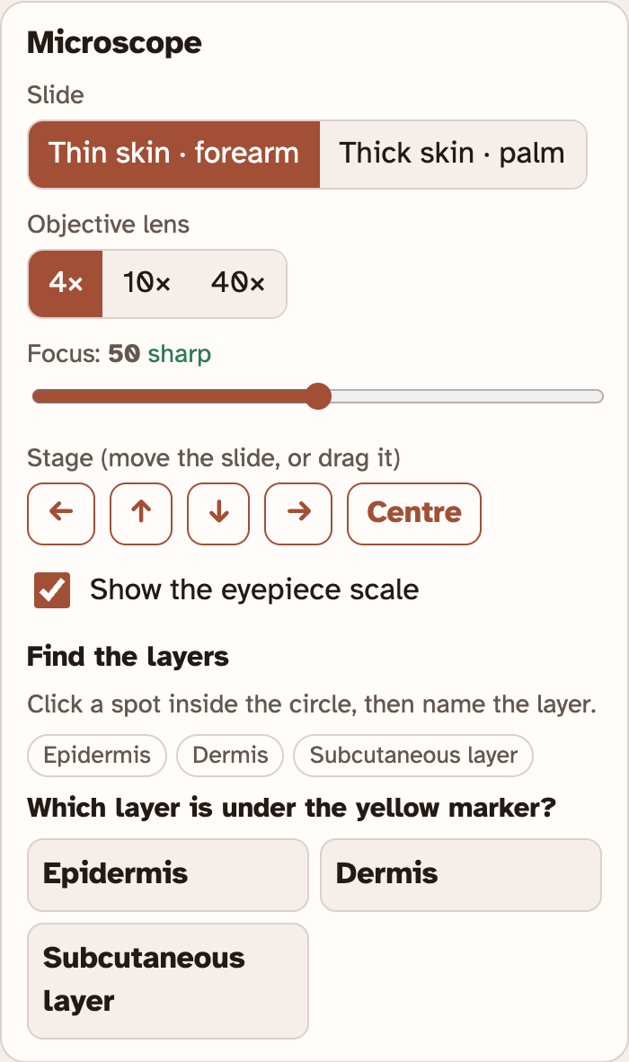
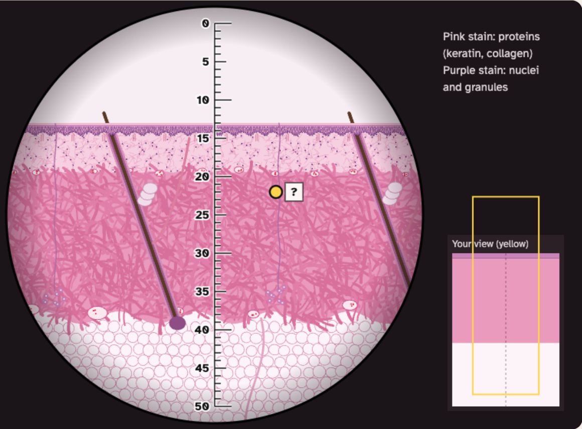
Drag the slide to move it. Or use the arrow buttons.
Pink stain shows proteins. Purple stain shows nuclei.
Screenshots: mrsajdak.com/skin-layers, Tab 1
PART 1 · QUESTION 2
Read the eyepiece scale
1
Find the scale in the middle of the circle. It has 50 divisions.
2
Pick the 4× objective lens. Look to the left of the circle. The lab says what 1 division is worth.
3
Write that number. Do the same for 10× and 40×.
4
Focus again after each change. Each lens has its own sharp spot.
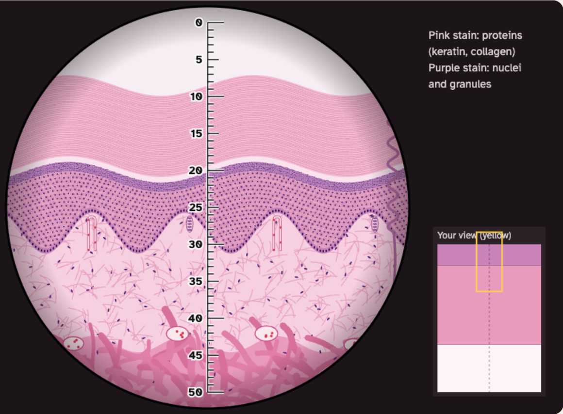
Words to know
Objective lens
The lens near the slide.
Division
One small step on the scale.
µm
A micrometer. 1,000 µm = 1 mm.
Screenshot: mrsajdak.com/skin-layers, Tab 1
PART 1 · QUESTION 3
Measure a layer
1
Press Centre. Your worksheet uses the middle of the slide.
2
Move the slide up or down. Put 0 on the top edge of the layer.
3
Count the divisions down to the bottom edge of the layer.
4
In Measure a layer, pick the layer. Type your count. Press Check.
5
Fill in the row. Do both layers on both slides.
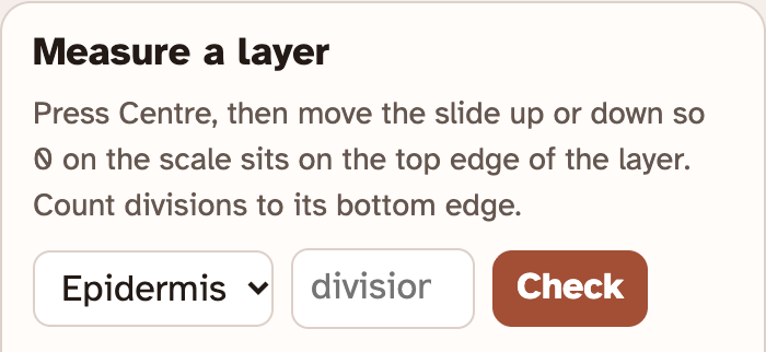
Layer longer than the scale? Try a weaker lens.
Layer only a division or two? Try a stronger lens.
The lab keeps your checked rows in Your results.
Example
You count 9 divisions.
One division is worth 25 µm.
9 × 25 = 225 µm
225 µm ÷ 1,000 = 0.225 mm
These are not your numbers. Use your own.
Screenshot: mrsajdak.com/skin-layers, Tab 1
PART 1 · QUESTION 4
How deep does it go?
1
Pick a slide.
2
Under How deep is the injury?, press Scrape · 0.05 mm.
3
Find the white notch on the slide. Read Deepest layer and Bleeds?
4
Do the same for Cut · 0.3 mm and Deep cut · 2.0 mm.
5
Switch slides. Do all three again.
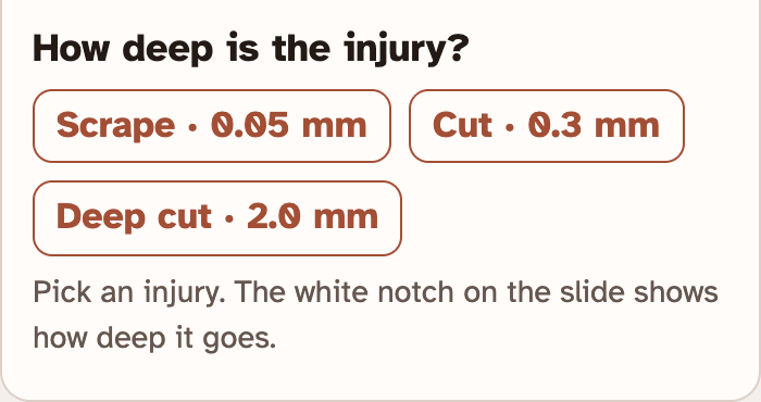
If the lab says Yes, a little, circle yes.
Your results keeps a list under Injuries.
Screenshot: mrsajdak.com/skin-layers, Tab 1
PART 1 · LEARN IT · 1 OF 4
Epidermis: the cover
Stacked cells that keep water in and germs out. It has no blood vessels. Food comes up from below.
Photo: Kilbad, public domain
PART 1 · LEARN IT · 2 OF 4
Dermis: the living support
Tough fibers, plus the skin's blood vessels, nerves, glands and hair roots. A cut that reaches it can bleed.
Screenshot: mrsajdak.com/skin-layers, Tab 1
PART 1 · LEARN IT · 3 OF 4
Subcutaneous layer
Mostly fat. It holds the skin to the muscle, keeps heat in and softens bumps. It is under the skin.
Screenshot: mrsajdak.com/skin-layers, Tab 1
PART 1 · LEARN IT · 4 OF 4
Pick the lens that fits
A layer that covers 1 or 2 divisions is hard to count. A stronger lens spreads it over more divisions.
Screenshot: mrsajdak.com/skin-layers, Tab 1
PART 1 · QUESTIONS 5 AND 6
Use the three jobs
QUESTION 5
Best?
Which lens for the thin epidermis?
Look at your table. Which objective did you use?
How many divisions did it cover at 4×? Use that to explain.
QUESTION 6
0.3 mm
Same cut, two results
Compare the epidermis on the forearm and on the palm. Use your Question 3 numbers.
Use the words blood vessels.
DAY 1 · TAB 2 · QUESTIONS 7 TO 10
Part 2. Body map
Skin is not the same thickness everywhere.
You will compare six places on the body. Look for a pattern.
Photo: Western Arctic National Parklands, CC BY 2.0
PART 2 · QUESTION 7
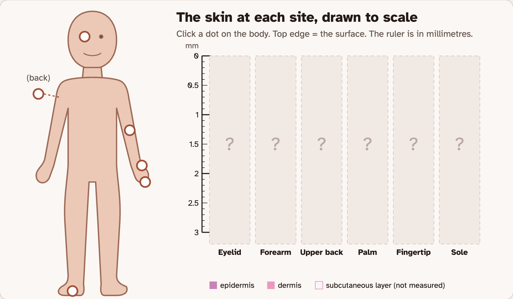
Click all six sites
1
Click each of the six dots on the body.
2
Each click adds a slice of that skin to the chart. Every slice is drawn to the same scale.
3
Find the Six sites table.
4
Copy Epidermis mm and Dermis mm for each site.
5
The Skin column says thick or thin. Copy that too.
Screenshot: mrsajdak.com/skin-layers, Tab 2
PART 2 · QUESTION 8
Line them up
QUESTION 8
÷
How many times thicker?
Divide the sole epidermis by the eyelid epidermis.
Then find the forearm in your Question 3 table. Does it match the chart?
SORT 1
Epidermis
Thin to thick
Under Line the slices up by, press epidermis, thin to thick.
Click the three thickest. Read What it faces for each one.
SORT 2
Dermis
Thin to thick
Press dermis, thin to thick. Find the upper back.
Read What it faces for the back.
PART 2 · LEARN IT · 1 OF 4
Thick skin, thin skin
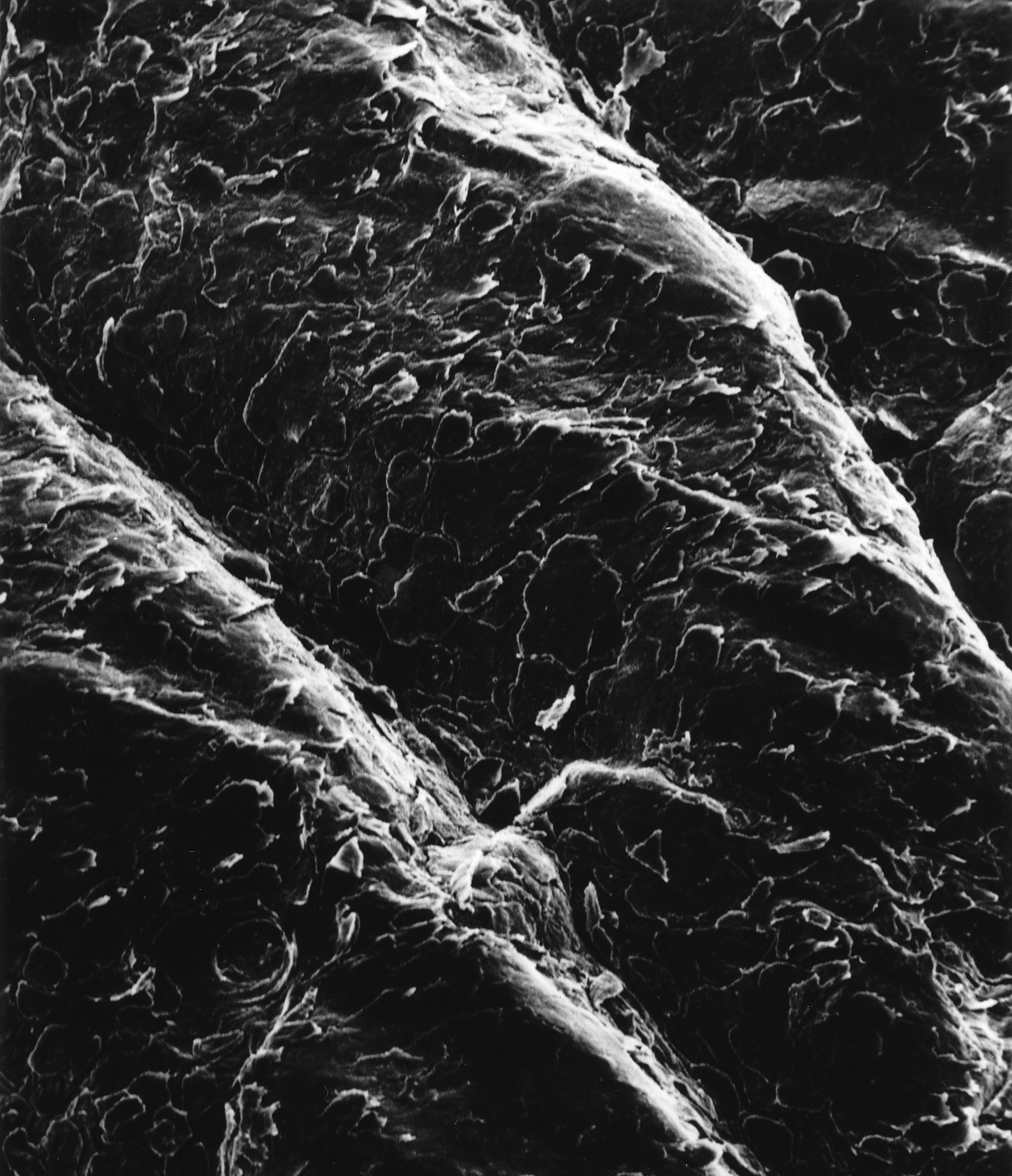
The names are about the epidermis only. Thick skin is on the palms, fingertips and soles. It has no hair.
Photo: Western Arctic National Parklands, CC BY 2.0
PART 2 · LEARN IT · 2 OF 4
The epidermis takes the wear
Its top is dead cells full of keratin. Rubbing wears them off first. A thick stack protects the living cells below.
Photo: Emily Hassell, National Park Service
PART 2 · LEARN IT · 3 OF 4
The dermis takes the pull
Collagen fibers are strong. Elastic fibers stretch and snap back. A thick dermis can be pulled without tearing.
Screenshot: mrsajdak.com/skin-layers, Tab 4
PART 2 · LEARN IT · 4 OF 4
Structure fits function
Each place has more of the layer whose job it needs most.
Screenshot: mrsajdak.com/skin-layers, Tab 2
PART 2 · QUESTIONS 9 AND 10
Find the pattern
QUESTION 9
Top 3
What do they share?
Look at the three thickest epidermis slices. What do those places do all day?
Why does that need a thick epidermis?
QUESTION 10
Back
Thick dermis, thin epidermis
The back is pulled and bent, but almost never rubbed.
Use the job of each layer to explain.
DAY 2 · TAB 3 · QUESTIONS 11 TO 15
Part 3. Inside the epidermis
The epidermis has layers of its own. One layer is a stratum. More than one are strata.
You will name them. Then you will follow one cell from the day it is born to the day it flakes off.
Photo: Bruce Wetzel, National Cancer Institute
PART 3 · QUESTION 11
Name the five strata
1
Stay on Thick skin · palm. Click a band in the picture.
2
Name it. A wrong pick gets crossed out. Try again.
3
Read the note that shows up. It tells what happens to cells there.
4
Find all five. Write them in order, bottom to top.
5
Switch to Thin skin · forearm. Which stratum is missing?
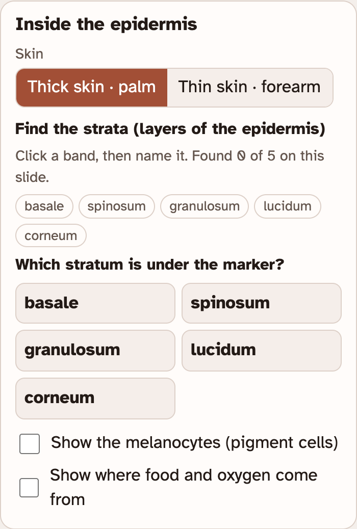
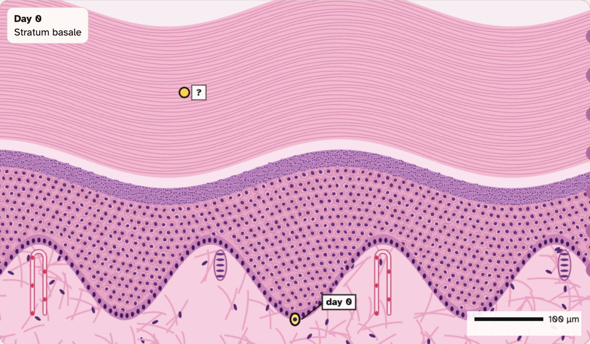
The yellow marker shows where you clicked.
Found bands get a green pin with their name.
Screenshots: mrsajdak.com/skin-layers, Tab 3
PART 3 · QUESTION 12
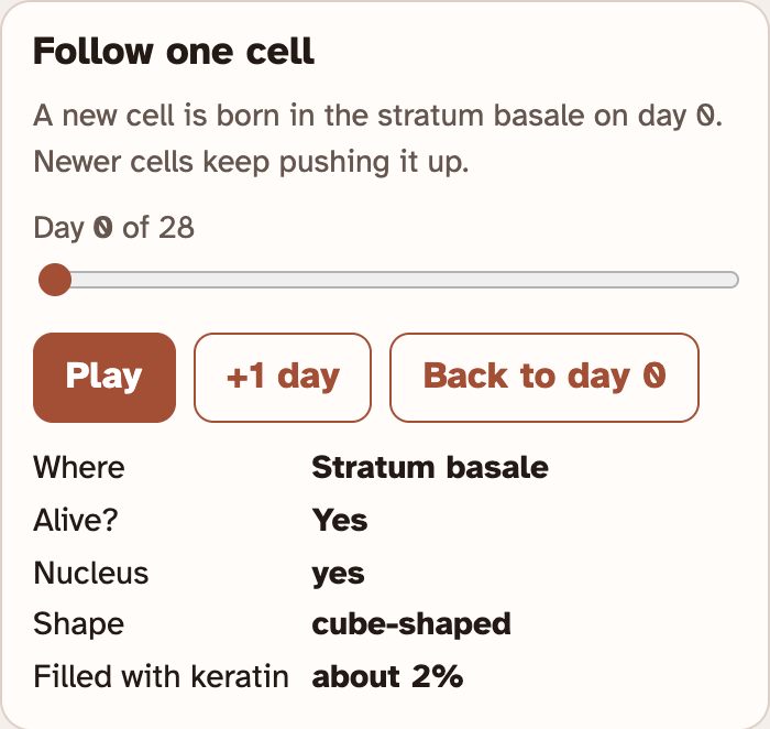
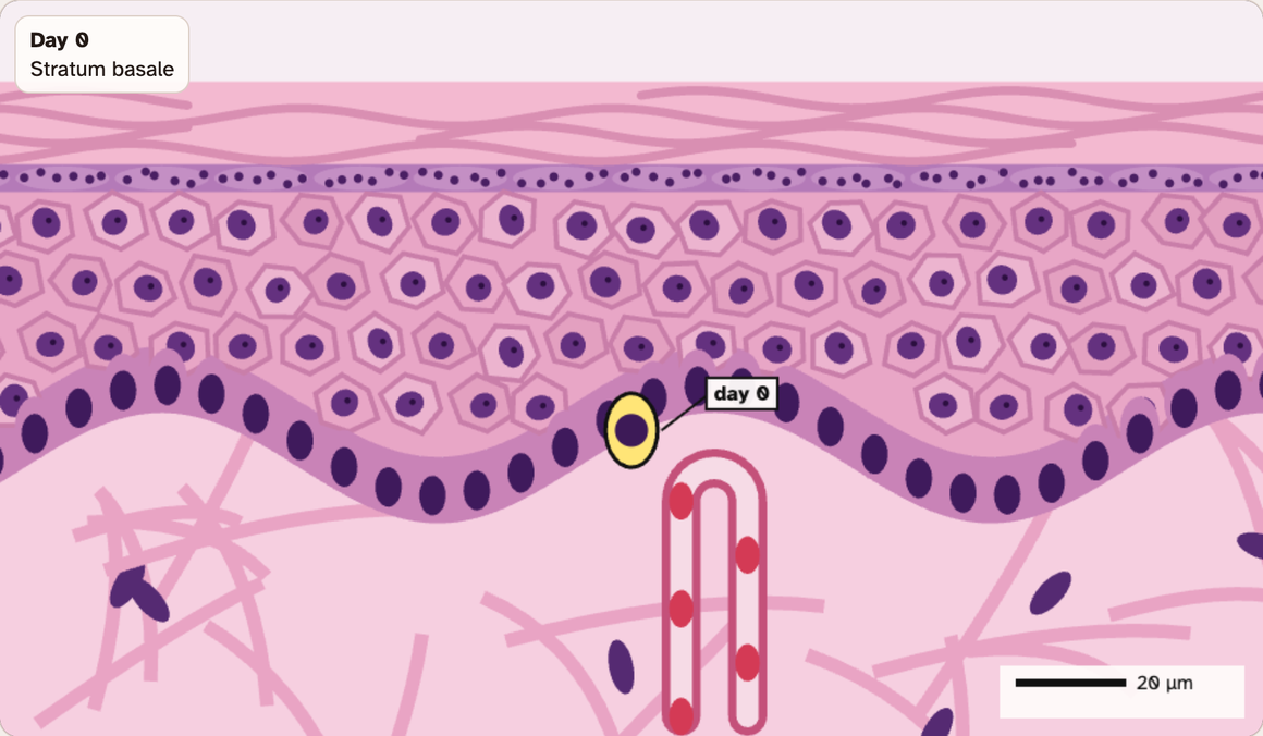
Follow one cell
1
Switch to Thin skin · forearm.
2
Find Follow one cell. Press Play. Or press +1 day.
3
Watch The cell’s trip. It fills in the day the cell enters each stratum.
4
Copy the days. The last one is the day the cell flakes off.
5
Divide that last day by 7. That is the number of weeks.
Example
42 ÷ 7 = 6
A cell that flakes off on day 42 took 6 weeks. These are not your numbers.
Screenshots: mrsajdak.com/skin-layers, Tab 3
PART 3 · QUESTION 13
Where does the cell die?
QUESTION 13
Alive?
Where does the cell die?
Step through the days. Watch Nucleus. Where does it start to break down?
Check your notes from Question 11. Then read the last line in the box. What fills the cell?
LOOK
Glow
Where is the food?
Tick Show where food and oxygen come from. Find the yellow glow.
Where does it start? Which cells are far from it?
PART 3 · LEARN IT · 1 OF 4
Fed from below
No blood vessels here. Food and oxygen leave the dermis and spread up by diffusion: from a lot to less.
Screenshot: mrsajdak.com/skin-layers, Tab 3
PART 3 · LEARN IT · 2 OF 4
Near and far
Cells at the bottom are close to the supply. They live and divide. New cells push older ones up, farther away.
Screenshot: mrsajdak.com/skin-layers, Tab 3
PART 3 · LEARN IT · 3 OF 4
Keratinization
On the way up, a cell fills with keratin, its nucleus breaks down and it dies. Dead cells make a tough cover.
Photo: Bruce Wetzel, National Cancer Institute
PART 3 · LEARN IT · 4 OF 4
In equals out
A tub with the tap on and the drain open stays level if water comes in as fast as it leaves. So does a layer of cells.
Screenshot: mrsajdak.com/skin-layers, Tab 3
PART 3 · QUESTIONS 14 AND 15
Fed and replaced
QUESTION 14
Fed?
No blood vessels
Use the word diffusion to explain how the epidermis is fed.
The top cells are far from the food. Why is it fine that they are dead?
QUESTION 15
Steady
Lost on top, same thickness
Cells leave the top every day.
Which stratum keeps up with the loss? How?
DAY 2 · TAB 4 · QUESTIONS 16 TO 19
Part 4. Inside the dermis
The dermis has two layers. The papillary layer is on top. The reticular layer is below.
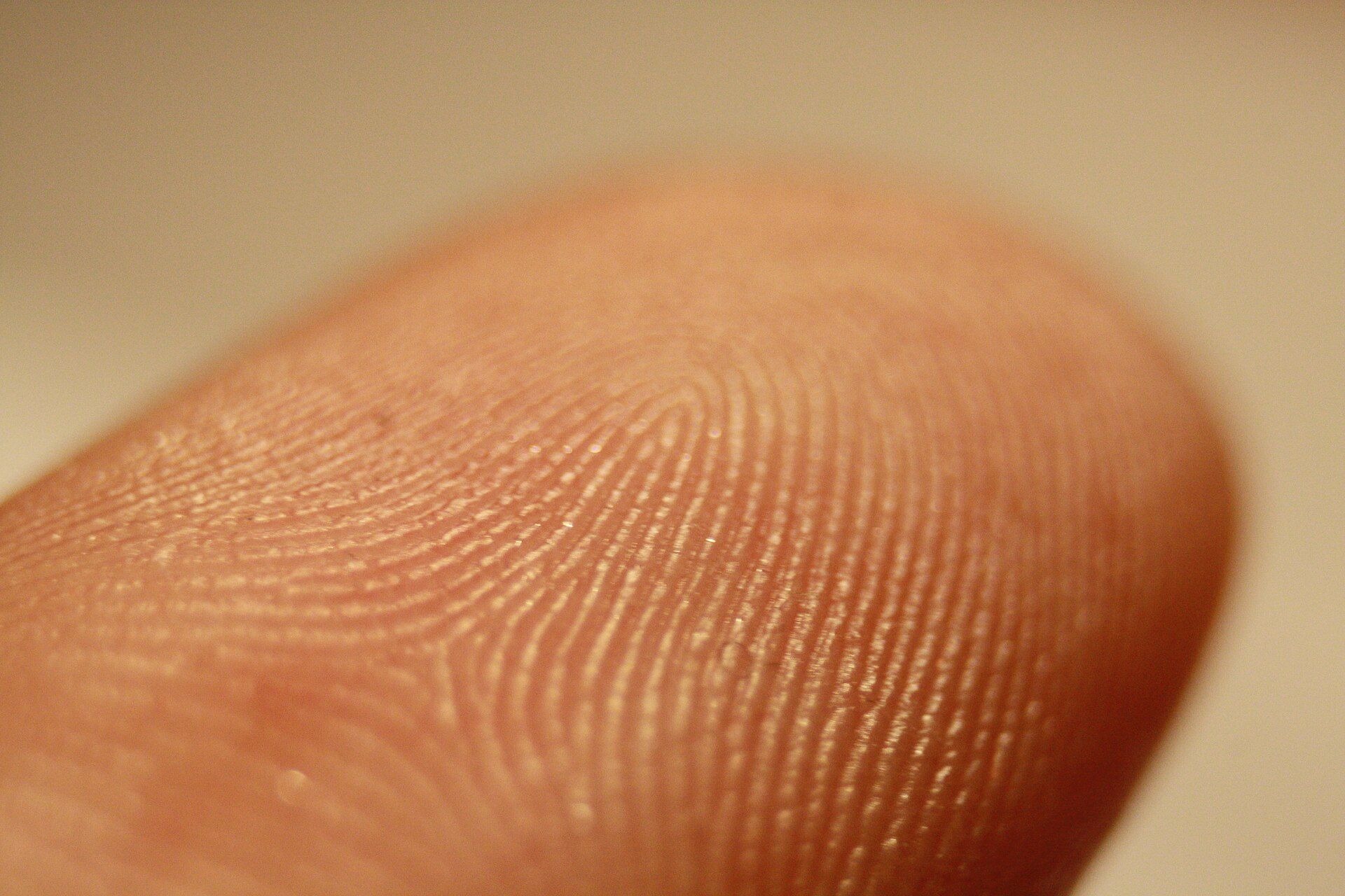
You will find seven structures, pull on each layer, and count the ridges on a fingertip.
Photo: Frettie, CC BY 3.0
PART 4 · QUESTION 16
Find all seven
1
Look under Find each one. The lab names what to find next.
2
Click it inside the circle. Green words tell you when you found it.
3
Wrong spot? The lab tells you what you clicked. Keep looking.
4
Move the slide down for the deep ones. Or switch to 4×.
5
Read the note under each one you find. It says where it is. Then tick the table.
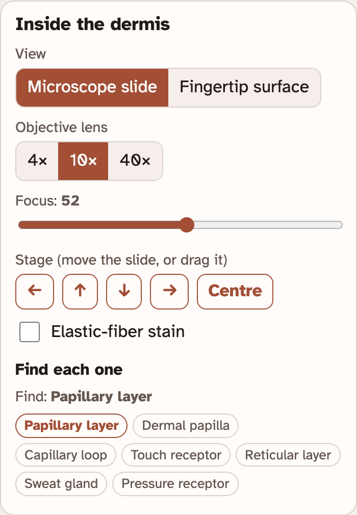
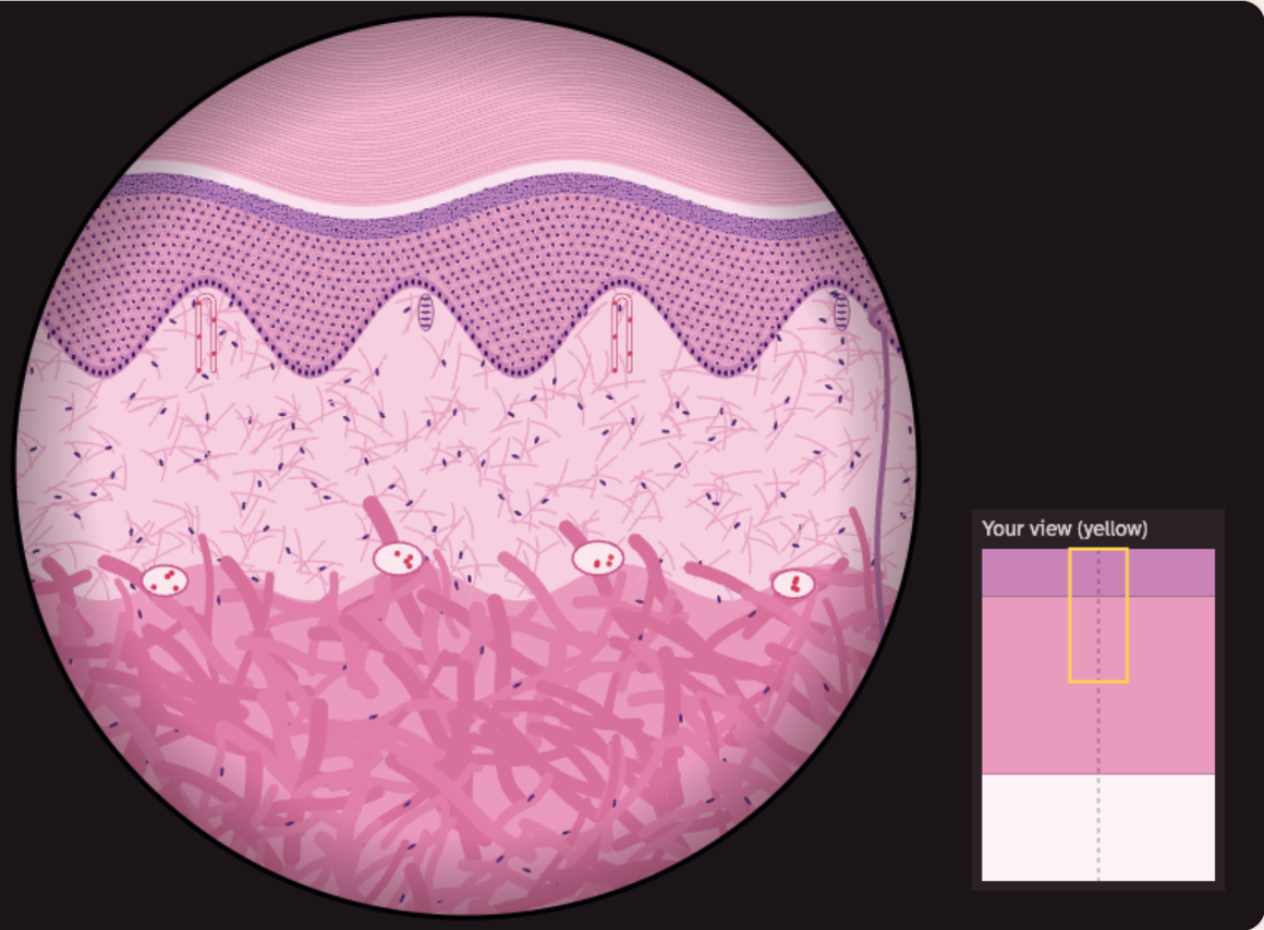
You find them in order. There are seven.
Elastic-fiber stain turns the elastic fibers black.
Screenshots: mrsajdak.com/skin-layers, Tab 4
PART 4 · QUESTIONS 17 AND 18
Pull until it tears
1
Find Pull test. Pick a Sample and a Pull direction.
2
Press Skip to the end. Or press Pull until it tears and watch.
3
Read the force where it tore. It is in Your results.
4
Do all five tests in your table.
5
Question 18: divide the reticular force by the papillary force.
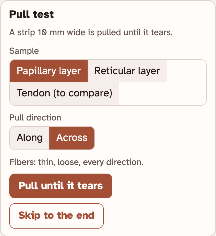
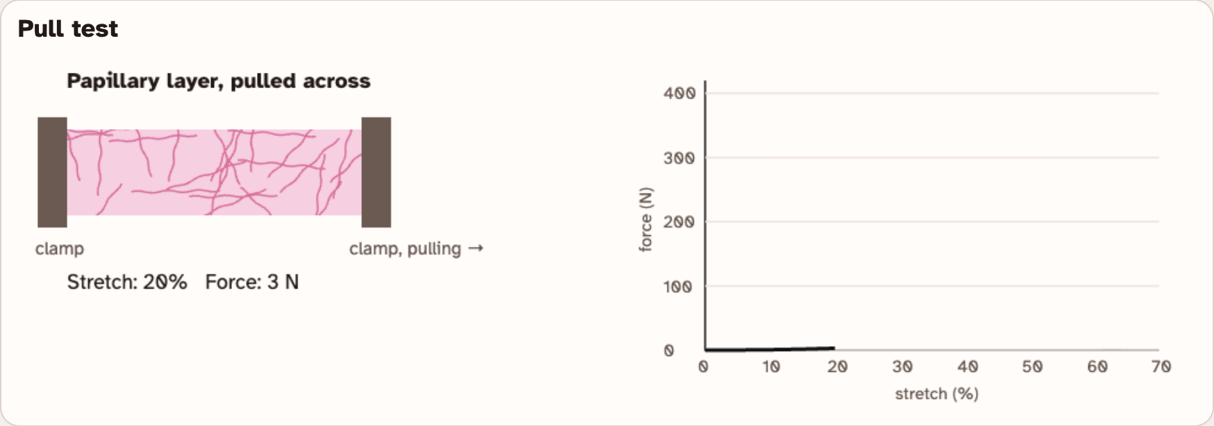
This strip is still being pulled. It has not torn yet.
Read Fibers under the buttons. It says how the fibers run.
Example
60 ÷ 15 = 4
A strip that tears at 60 N is 4 times stronger than one that tears at 15 N. These are not your numbers.
Screenshots: mrsajdak.com/skin-layers, Tab 4
PART 4 · QUESTION 18
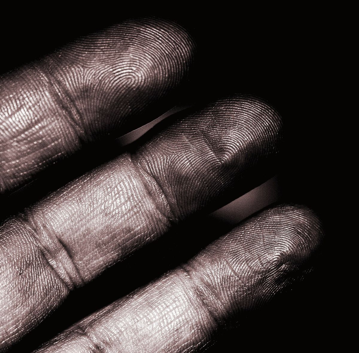
Count the ridges
1
Under View, press Fingertip surface.
2
Find the blue line inside the 5 mm box. Count the ridges that cross it.
3
Divide 5 mm by your count. That is how far apart the ridges are.
4
Write both numbers in Question 18.
Example
5 mm ÷ 4 = 1.25 mm
4 ridges across 5 mm are 1.25 mm apart.
These are not your numbers.
Screenshot: mrsajdak.com/skin-layers, Tab 4 · Photo: D. Sharon Pruitt, CC BY 2.0
PART 4 · LEARN IT · 1 OF 4
Fibers carry the pull
A collagen fiber is strong when pulled along its length. Pulled from the side, fibers slide apart.
Screenshot: mrsajdak.com/skin-layers, Tab 4
PART 4 · LEARN IT · 2 OF 4
Two ways to line them up
Dense regular: all one way, like a tendon. Dense irregular: bundles in every direction, like the reticular layer.
Screenshot: mrsajdak.com/skin-layers, Tab 4
PART 4 · LEARN IT · 3 OF 4
Papillae make the ridges
Rows of dermal papillae push up into the epidermis. The epidermis forms over them as friction ridges.
Photo: Frettie, CC BY 3.0
PART 4 · LEARN IT · 4 OF 4
The dermis stays
The epidermis is replaced every few weeks. The dermis is never shed. Its fibers are kept and repaired in place.
Photo: D. Sharon Pruitt, CC BY 2.0
PART 4 · QUESTION 19
Why it is built that way
QUESTION 19a
Strong both ways?
Find your reticular and tendon numbers from Question 17, along and across.
Why is the reticular layer strong both ways, but a tendon only one way? Use how the fibers are arranged.
QUESTION 19b
Fingerprints for life
The epidermis is replaced every few weeks.
So why are your fingerprints the same all your life?
WRAP IT UP · QUESTION 20
Why does a tattoo last?
The claim
“Tattoo ink lasts for life only because it is put into the dermis.”
Evidence 1
Pick one tab. Use a number or a fact from your worksheet. Tab 3 tells how long one cell lasts.
Evidence 2
Pick a different tab. Tab 1 shows how deep a cut must go. Tab 4 shows a pattern that lasts for life.
Reasoning
Compare the two layers. What happens to cells in the epidermis over time? What about the dermis?
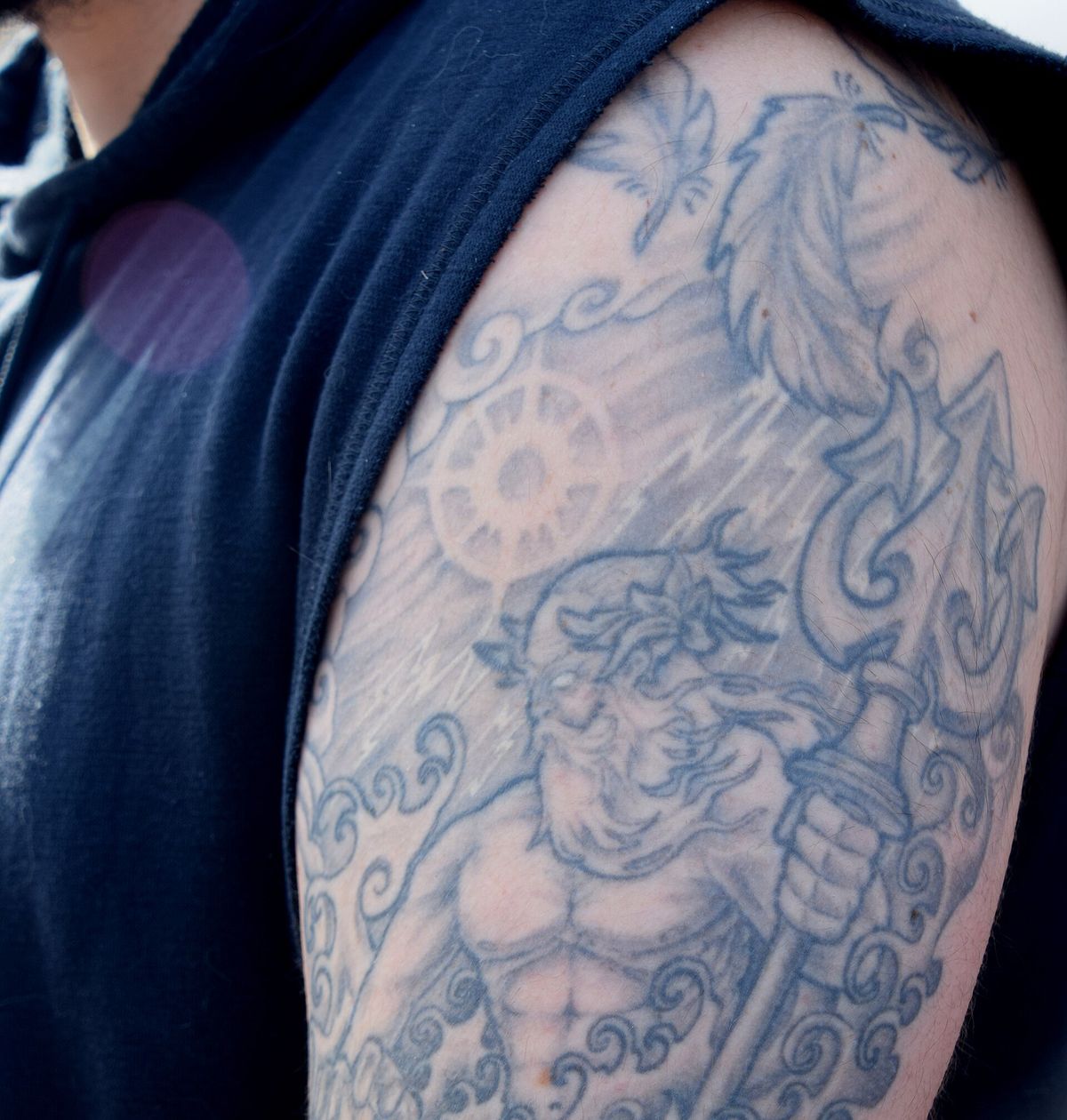
Tattoo ink sits in the skin for many years.
Photo: Tony Alter, CC BY 2.0
WRAP IT UP · BACK TO THE START
Revisiting the Phenomenon
Why does a tattoo stay when skin wears away?
1Using evidence from Part 3, explain what happens to a cell in the epidermis, from the day it is made to the day it flakes off.
2A tattoo needle puts ink into the dermis. Use your numbers from Question 3. About how deep must the needle go on the forearm?
3Why does ink in the dermis stay, when ink in the epidermis would be gone in weeks? Use the names of both layers.
4Fingerprint ridges also last for life (Question 19). How is a fingerprint like a tattoo?
LAST CHECK
Before you hand it in
Name, date and period are on top.
Every table is full, front and back.
Questions 1, 11 and 16 have their ticks.
Every measurement has a unit: µm, mm or N.
Question 20 uses evidence from two different tabs.
Photo: R G, CC BY 2.0