Solid or Spongy? Bone Tissue Lab
A real human thigh bone, cut open. Measure its walls, drop water on it, look at it under a microscope, and find out why some parts of the skeleton break first when bones get weak.
Graph
Learn It: two kinds of bone tissue
Finish these steps in the lab. Then this box opens.
The parts of a long bone
The long middle of a bone like the femur is the diaphysis (the shaft). Each end is an epiphysis. The empty channel in the shaft is the medullary cavity. In a living person it is full of soft marrow.
Compact bone
The thick, solid wall of the shaft is compact bone. It has almost no open space. You measured it at about 7 mm. A thick, solid tube is hard to bend or twist. That is its job.
Spongy bone
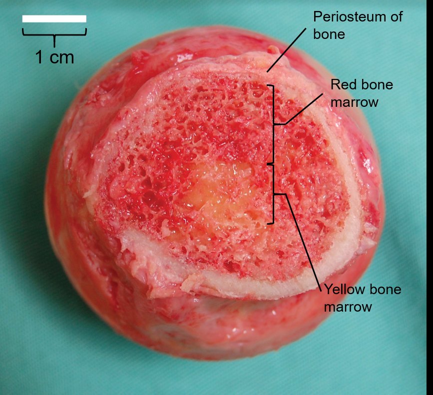
The inside of the end is spongy bone. It is a lattice of thin, hard plates with spaces between them. Only a thin shell of compact bone covers it. Yours was about 3 mm.
Spongy does not mean soft
Spongy bone is made of the same hard material as compact bone. It is built as a lattice instead of a block. "Spongy" is about the holes, not the feel. The holes make the end lighter. The plates line up with the pushes that come from the joint.
Why the water vanished
On the solid wall, the drop sat on top. On the spongy end, it soaked in within seconds. The water ran into the open spaces. In a living bone those spaces are full of red marrow, which makes blood cells.
Now answer Q6 to Q7 on your worksheet.
Learn It: what the microscope shows
Finish these steps in the lab. Then this box opens.
Osteons
Compact bone is built of osteons: rings of hard bone matrix around a tube. Each ring is a thin layer. You measured one osteon at about 170 µm across. That is about the width of two hairs. An osteon is long, like a pencil, and runs along the shaft.
The central canal
The tube in the middle of each osteon is the central canal. Yours was about 50 µm across. It carries blood vessels and nerves, so the bone stays alive.
Bone cells
The dark specks between the rings are tiny spaces. Each one holds a bone cell, an osteocyte. Thin channels link the cells to each other and to the canal. That is how food reaches cells inside hard rock-like matrix.
Trabeculae
The purple plates in the spongy bone picture are trabeculae. They have no osteons. They are thin enough to get food from the marrow around them. Between them is marrow: big white fat cells and small blue blood-making cells.
Your counts
In compact bone, 24 of 25 points landed on hard bone. Only about 4% was open space. In spongy bone, only 10 of 25 points did. About 60% was open space. Real measurements agree: compact bone is usually less than 5% open space, and in spongy bone only about 20% of the space is bone.
Structure fits function
Same hard material, two ways to build it. Packed tight, it makes a strong tube for the shaft. Built as a lattice, it makes a light end that holds marrow and spreads the push from a joint.
Now answer Q11 to Q13 on your worksheet.
Learn It: osteoporosis
Finish these steps in the lab. Then this box opens.
What osteoporosis is
Osteoporosis means "porous bone." Bone is living tissue. Cells take old bone away and build new bone all the time. With age, more is taken away than is built. The bone gets lighter and full of bigger holes, and it breaks more easily.
Spongy bone goes first
Spongy bone has much more surface touching the marrow, and that is where bone cells work. So it changes fast. In a large study, people lost spongy bone from young adulthood on. Compact bone loss started later, in middle age. Plates get thin, then break, and the lattice loses its links.
The model
The lab mixed two real numbers. First, how much of each site is compact or spongy. Second, how much of each tissue a person loses over a lifetime. Sites with the most spongy bone lost the most. The spine is about 75% spongy. The shaft of the forearm bone is only 5% spongy.
Hip, spine and wrist
About 1 in 2 women and 1 in 4 men over 50 will break a bone because of osteoporosis, most often at the hip, spine or wrist. All three are spongy-rich places. The hip break is at the top of the femur, an end. The spine is made of vertebrae, spongy inside. The wrist break is at the end of the radius, not its shaft.
Women and men
Women lost more bone in the study than men, and they start with smaller bones. That is a big reason hip breaks are more common in older women.
Now answer Q16 to Q17 on your worksheet.
Printable student worksheet (PDF) · two pages, goes with this lab
Low-write worksheet (PDF) · same questions with tick boxes, word banks and worked examples
Teacher guide: NYSSLS, SEP and CCC alignment (PDF)
Slideshow for the board
What you will learn. I can compare where compact and spongy bone are and what each one looks like. I can explain how the structure of each tissue fits its job. I can use bone data to explain why osteoporosis breaks some bones first.
Photos. Tab 1: a human femur cut across its shaft (MAKY.OREL, Charles University anthropological collection, CC0) and the neck of a human femur cut across after hip replacement surgery (Steven Fruitsmaak, public domain, with a 1 cm bar). The shaft photo has no scale bar, so its scale is set from a typical adult femur, about 30 mm across at this cut: its readings are close, not exact. The drop times are a model, set to match a clean, dry bone; a real bone can be faster or slower. Tab 2: a ground section of compact bone (Lord of Konrad, CC0; scale from the 40 µm bar on Katsamenis's black-and-white copy of the same photo, public domain) and spongy bone from a human vertebra (Nataliia Maryenko, CC BY-SA 4.0, scale from her 100 µm bar). Tab 3: slices of a healthy vertebra (a 37-year-old man) and an osteoporotic one (a 75-year-old woman), Turner Biomechanics Laboratory, CC0. Learn It: the end of a humerus, OpenStax, CC BY 4.0. Numbers: compact bone porosity under 5% and the compact-to-spongy ratios of the radius shaft (95:5), femoral head (50:50) and vertebra (25:75) from Clarke B (2008), Normal bone anatomy and physiology, Clin J Am Soc Nephrol 3: S131–S139; spongy bone about 20% bone by volume from Ott SM (2018), Am J Nephrol 47: 373–375. Bone lost over life (spongy bone density at the spine and hip down about 55% in women and 46% in men; compact bone density down about 25% and 18%) from Riggs BL and others (2004), J Bone Miner Res 19: 1945–1954, with spongy loss starting in young adults and compact loss in midlife from Riggs and others (2008), J Bone Miner Res 23: 205–214. The lab spreads each loss evenly over the years (spongy from 20 to 90, compact from 50 to 90): a simplification. Fracture numbers: NIH. Hole's Essentials of Human Anatomy & Physiology, Section 7.2.
© 2026 Ryan Sajdak. This lab, its worksheets and its slides are free to use, share and change under the Creative Commons BY-NC-SA 4.0 license: give credit, do not sell it, and share your version the same way. Photos keep their own licenses, listed above.