ANATOMY & PHYSIOLOGY · SECTION 7.2
Solid or Spongy?
Bone tissue lab. Follow along with your worksheet, one question at a time.
mrsajdak.com/bonetissue
Photo: Steven Fruitsmaak, public domain
START HERE · PICK AN OPENER
Choose how to open the lesson

Click a card to jump to it. Every option ends at the same Observe, Wonder, Predict slide and the same driving question.
OPTION A · VIDEO
Cut Through a Bone
Clip 1 is a butcher cutting a beef marrow bone on a band saw. Clip 2 is a micro-CT scan, a 3D X-ray, of the inside of a bone. Clip 3 is a short news story about a new way to see bone loss.
Watch for
In Clip 1, look at the cut face. What is in the middle? What is around it?
In Clip 2, look at the inside of the bone. Is it solid?
Is every part of a bone built the same way?
Videos on YouTube: ASM butchers; Micro Photonics; Canadian Light Source.
OPTION B · TRY IT · 4 MINUTES
Sponge or Bone?
1
Put on gloves. Look at the cut end of the class bone. It is full of holes, like a sponge.
2
Press a kitchen sponge with a pencil eraser. Watch it squash.
3
Press the spongy end of the bone the same way. Tap it with the pencil.
4
Put one drop of water on the sponge and one on the bone end. Where does it go?
You need
A clean, dry kitchen sponge
The class bone, split, with a spongy end (cooked and cleaned)
A pencil
A dropper and a cup of water
Gloves
Watch for
The sponge squashes. The bone does not.
The water vanishes into both.
So what do the holes in bone do, if they do not make it soft?
OPTION C · PHOTOS
Phenomenon: One Bone, Two Cuts
Photos: MAKY.OREL, Charles University, CC0 · Steven Fruitsmaak, public domain
START HERE
Observe, wonder, predict
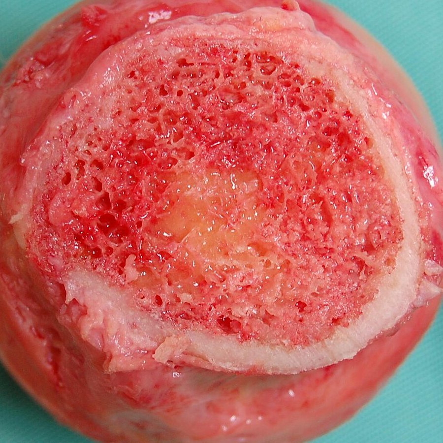
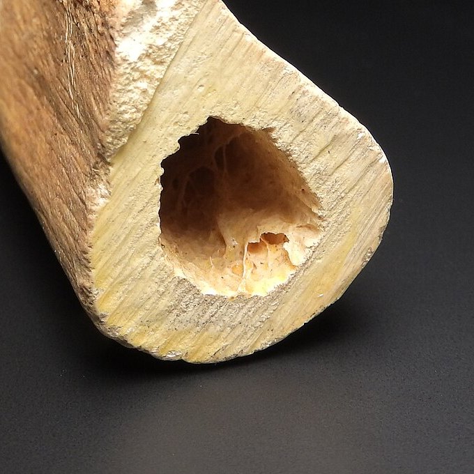
Both cuts are through a human thigh bone, the femur. One cut is solid all the way around a hole. The other is full of tiny holes, with only a thin rim.
OBSERVEWhat is different about the two cut faces?
WONDERWhat do you wonder about the holes?
PREDICTWhich part of the bone is stronger? Which is lighter? Explain your reasoning.
Cut A, up close.
Cut B, up close.
Photos: MAKY.OREL, Charles University, CC0 · Steven Fruitsmaak, public domain
DRIVING QUESTION
Why is one bone built two ways: solid in the middle, full of holes at the ends?
START HERE
Where Were These Cuts?
Finished observing? Click here to find out.
Cut A goes through the shaft of a femur, the long middle of the thigh bone. It is from a university bone collection. Cut B goes through the neck of a femur, just below the ball that fits into the hip. A surgeon removed it during a hip replacement. The red is marrow and blood.
Watched the video or tried it? Click here.
The butcher cut through a beef leg bone, and the 3D scan looked inside a bone. The kitchen sponge squashed, but the bone end did not, even though both are full of holes.
Today, you will measure the solid wall of a real femur and drop water on it. You will count points on real microscope pictures of bone. Then you will use real data to see which bones break first when bone gets weak. At the end, you will return to the driving question.
BEFORE YOU START
What you will learn
I can compare where compact and spongy bone are and what each one looks like.
I can explain how the structure of each tissue fits its job.
I can use bone data to explain why osteoporosis breaks some bones first.
BEFORE YOU START
Get set up
1
Open mrsajdak.com/bonetissue on your device.
2
Write your name, the date and your period on the worksheet.
3
Find the three tabs at the top of the lab. Each tab goes with one part of the worksheet.
4
Start on Tab 1. Do not skip ahead.

| Tab in the lab | Worksheet | Questions |
|---|---|---|
| 1. Cut the bone open | Part 1 | 1 to 7 |
| 2. Under the microscope | Part 2 | 8 to 13 |
| 3. Bone loss over a lifetime | Part 3 and Explain it | 14 to 17 |
Screenshot: mrsajdak.com/bonetissue
TAB 1 · QUESTIONS 1 TO 7
Part 1. Cut the bone open
Two cuts through a human femur.
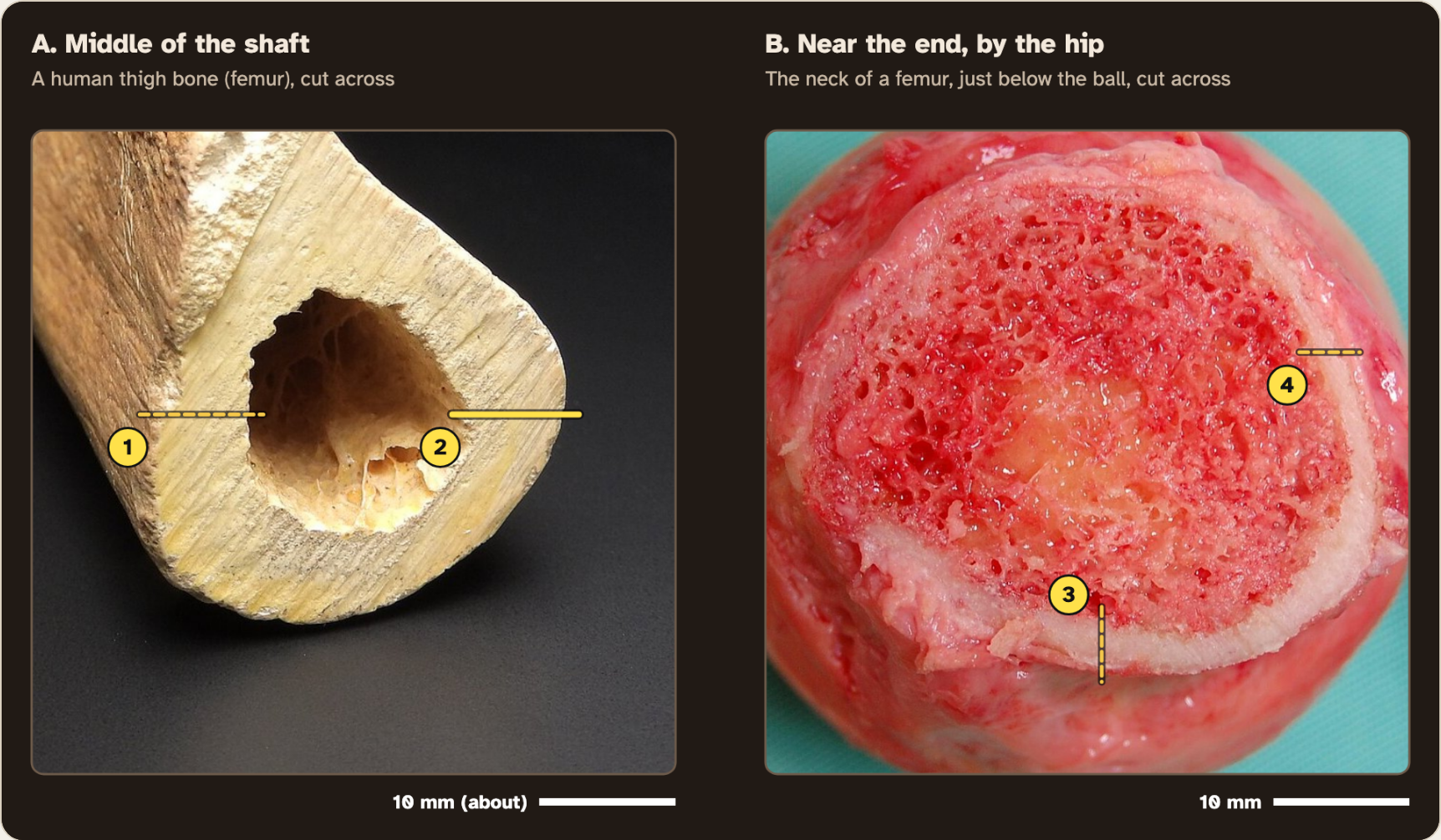
Measure the solid wall in the middle and near the end. Then drop water on each.
Photo: MAKY.OREL, Charles University, CC0
PART 1 · QUESTION 1
Look before you measure
Inside the end
Look at Cut B, in the middle of the red part.
Center of the shaft
Look at Cut A, in the middle.
Wall of the shaft
Look at Cut A, the pale ring.
For each place, tick one: solid and smooth, full of holes, or empty space.
Screenshot: mrsajdak.com/bonetissue, Tab 1
PART 1 · QUESTIONS 2 AND 3
Measure the solid wall
1
Click yellow spot 1. It shows up big in the Zoomed in box.
2
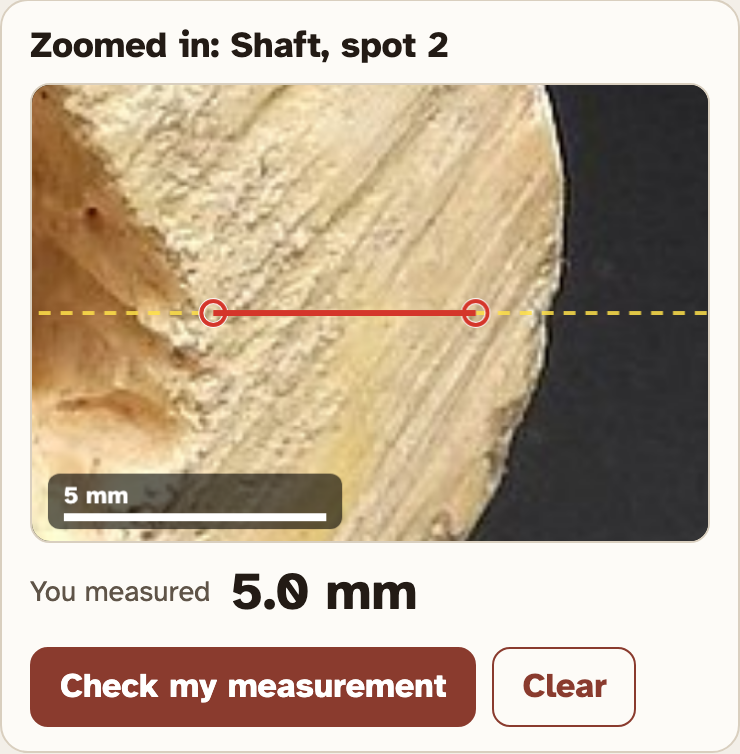
Click where the solid, pale wall starts. Then click where it ends.
3
Press Check my measurement. Write the lab’s number.
4
Do spots 2, 3 and 4.
PART 1 · QUESTIONS 2 AND 3
Average, then compare
QUESTION 2
Average
Add try 1 and try 2.
Divide by 2.
QUESTION 3
How many times as thick?
Shaft average ÷ end average.
Example
A wall is 9.0 mm in the middle and 3.0 mm near the end.
9.0 ÷ 3.0 = 3
These are not your numbers. Use your own.
The middle is 3 times as thick.
Screenshot: mrsajdak.com/bonetissue, Tab 1
PART 1 · QUESTIONS 4 AND 5
The drop test
1
Read Question 4 first. What changes? What is measured? What stays the same?
2
Press Drop on the solid wall. Watch the timer.
3
Press Drop on the spongy end.
4
Do each one twice. Write 60+ if the drop is still there at 60 s.
PART 1 · LEARN IT · 1 OF 4
The parts of a long bone
The long middle is the diaphysis (the shaft). Each end is an epiphysis. The empty channel in the shaft is the medullary cavity. In life it is full of soft marrow.
Photo: MAKY.OREL, Charles University, CC0
PART 1 · LEARN IT · 2 OF 4
Compact bone
The thick, solid wall of the shaft is compact bone. It has almost no open space. A thick, solid tube is hard to bend or twist. That is its job.
Photo: MAKY.OREL, Charles University, CC0
PART 1 · LEARN IT · 3 OF 4
Spongy bone
The inside of the end is spongy bone: a lattice of thin, hard plates with spaces between them. Only a thin shell of compact bone covers it.
Photo: Steven Fruitsmaak, public domain
PART 1 · LEARN IT · 4 OF 4
Why the water vanished
On the solid wall, the drop sat on top. On the spongy end, it soaked in within seconds. In a living bone, those spaces hold red marrow, which makes blood cells.
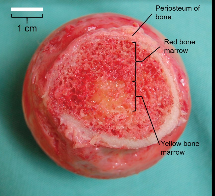
Photo: OpenStax, CC BY 4.0
PART 1 · QUESTIONS 6 AND 7
Name it
QUESTION 6
Wall · End · Channel
Three names
The solid wall of the shaft is ___ bone.
The inside of the end is ___ bone.
The empty channel is the ___.
QUESTION 7
60+ s vs 3 s
Where did the water go?
In a living bone, what fills those spaces?
What does it make?
TAB 2 · QUESTIONS 8 TO 13
Part 2. Under the microscope
The same two tissues, about 100 times bigger.
Count how much is hard bone. Then measure one osteon.
Photo: Lord of Konrad, CC0
PART 2 · QUESTION 8
Count the points
1
Pick Compact. Click every ring that sits on hard bone.
2
A ring inside a canal (the hole in the middle of a ring pattern) is open space. Dark specks count as bone.
3
Press Check my count. Then do Spongy: the purple plates are bone.
4
Each point is worth 4%, because 100 ÷ 25 = 4.
PART 2 · QUESTIONS 9 AND 10
Measure an osteon
1
Click the yellow O line, or the Osteon A button.
2
In the zoomed box, click one edge of the ring pattern, then the other.
3
Do the same for Canal A, the hole in its middle.
4
Write the lab’s numbers. 1,000 µm = 1 mm.
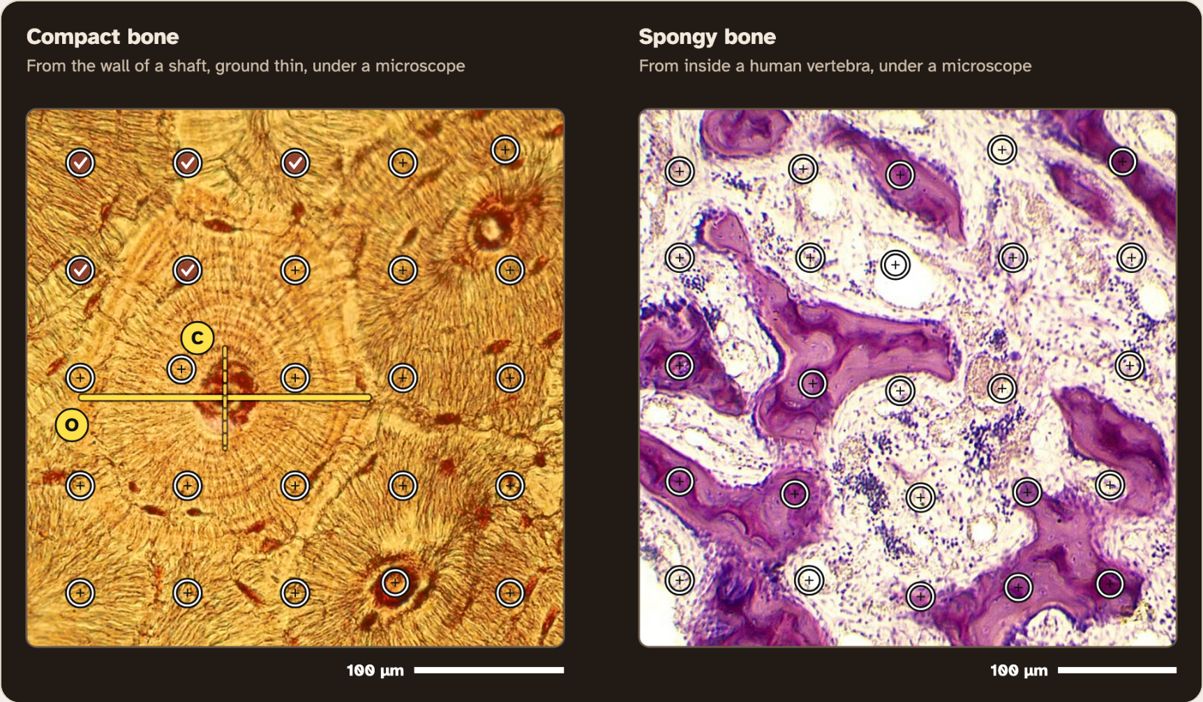
Screenshot: mrsajdak.com/bonetissue, Tab 2
PART 2 · LEARN IT · 1 OF 4
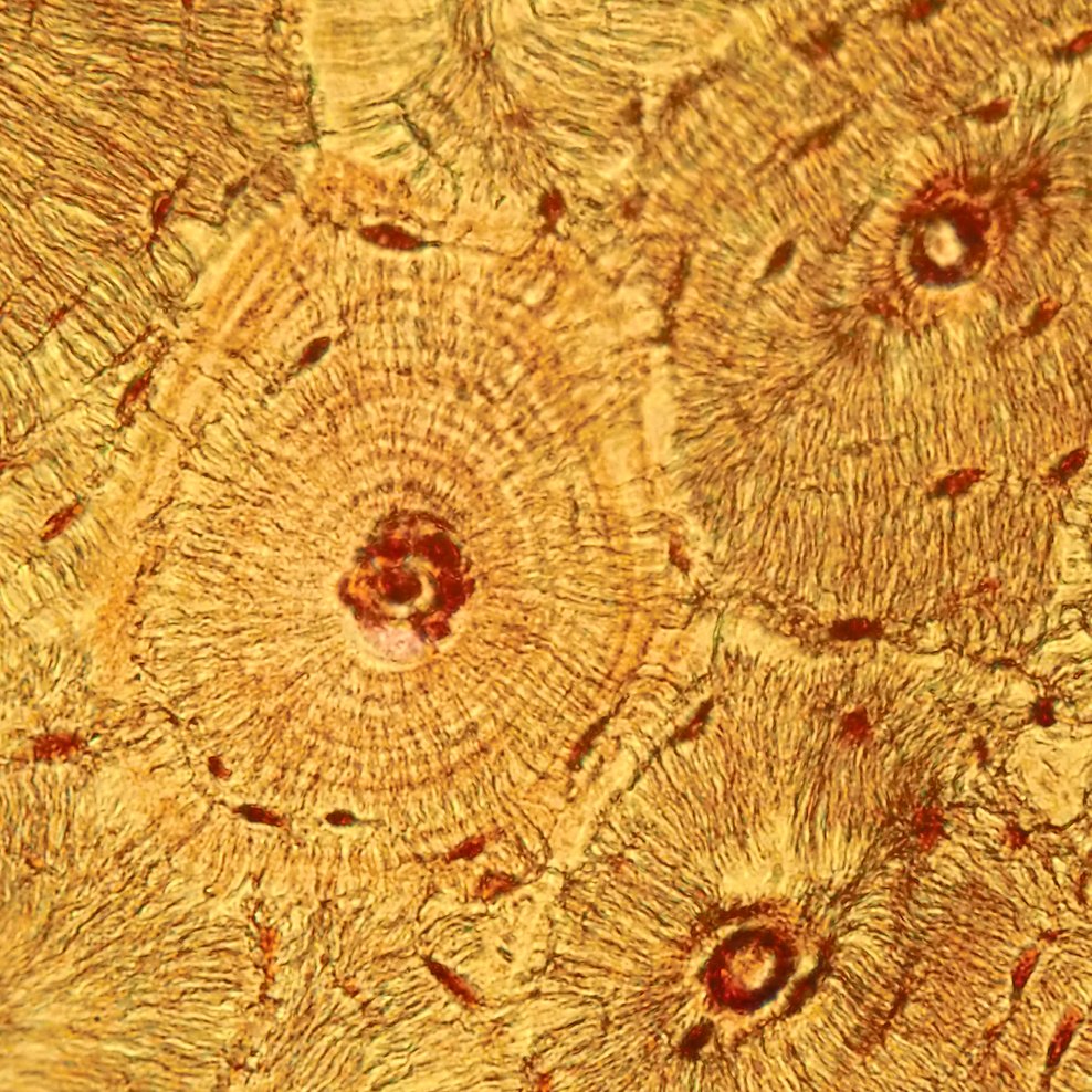
Osteons
Compact bone is built of osteons: rings of hard bone matrix around a tube. Each ring is a thin layer. One osteon is about as wide as two hairs, and long like a pencil.
Photo: Lord of Konrad, CC0
PART 2 · LEARN IT · 2 OF 4
Canal and cells
The tube in the middle is the central canal. It carries blood vessels and nerves. The dark specks are tiny spaces, each holding a bone cell, an osteocyte. Thin channels link them to the canal.
Photo: Lord of Konrad, CC0
PART 2 · LEARN IT · 3 OF 4
Trabeculae
The purple plates in spongy bone are trabeculae. Between them is marrow: big white fat cells and small blue blood-making cells.
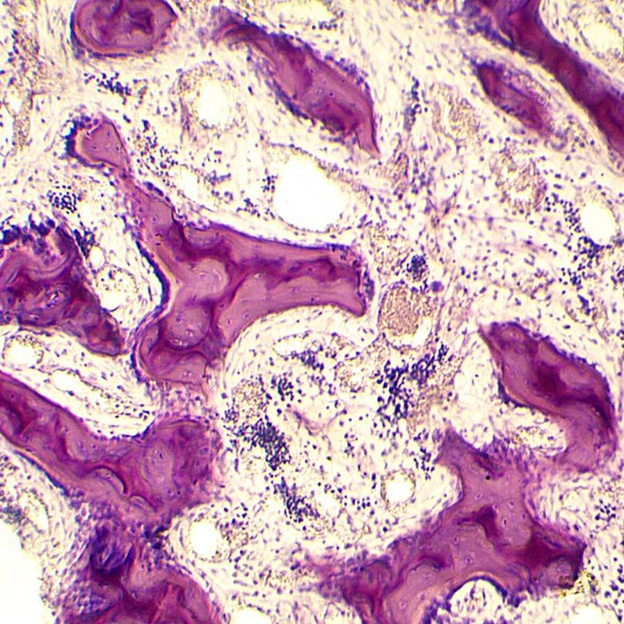
Photo: Nataliia Maryenko, CC BY-SA 4.0
PART 2 · LEARN IT · 4 OF 4
Spongy is not soft
Spongy bone is the same hard material as compact bone, built as a lattice instead of a block. Compact bone is less than 5% open space. In spongy bone, only about 20% of the space is bone.
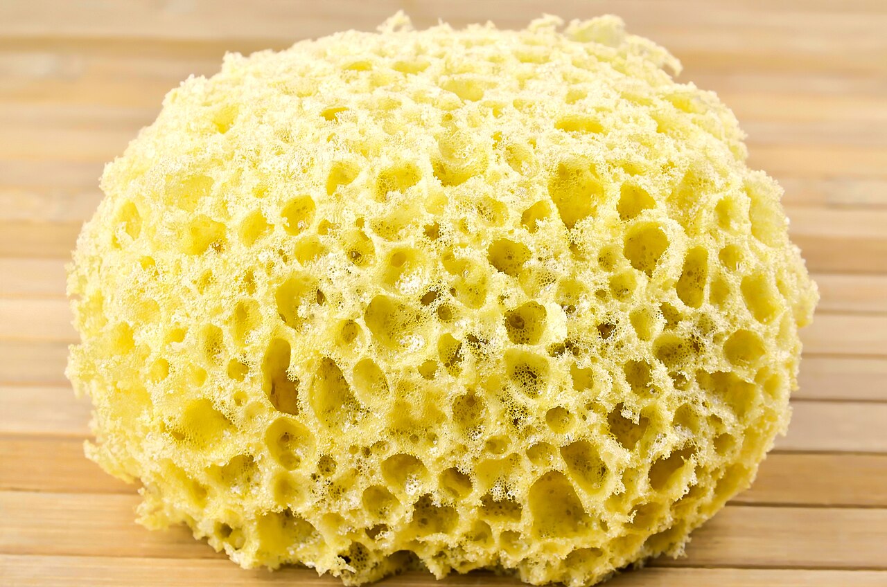
Photo: Robert Fotograf, CC0
PART 2 · QUESTIONS 12 AND 13
Use the idea
QUESTION 12
Soft?
"Spongy bone is soft, like a kitchen sponge."
Agree or disagree?
Give one piece of evidence from the lab.
QUESTION 13
Shaft · Ends
Structure and function
Why is compact bone in the shaft?
Why is spongy bone in the ends?
Name one job for each.
TAB 3 · QUESTIONS 14 TO 17
Part 3. Bone loss over a lifetime
Bone is alive. Cells take it away and build it up all the time.
Which parts of the skeleton lose the most bone with age?
Photo: Turner Biomechanics Laboratory, CC0
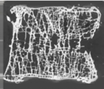
PART 3
Two vertebrae
Photos: Turner Biomechanics Laboratory, CC0
PART 3 · QUESTIONS 14 AND 15
Record three people
1
Pick Woman. Set the age to 50. Press Record this person.
2
Set the age to 90. Record.
3
Pick Man. Set 90. Record.
4
Which site lost the most? Look at how much of each site is spongy.
PART 3 · LEARN IT · 1 OF 3
Osteoporosis
Osteoporosis means "porous bone." With age, cells take away more bone than they build. The bone gets lighter, full of bigger holes, and breaks more easily.
Photo: Turner Biomechanics Laboratory, CC0
PART 3 · LEARN IT · 2 OF 3
Spongy bone goes first
Spongy bone has much more surface touching the marrow, where bone cells work. In a large study, people lost spongy bone from young adulthood on. Compact bone loss started in middle age.
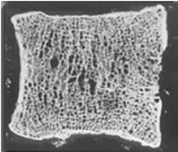
Photo: Turner Biomechanics Laboratory, CC0
PART 3 · LEARN IT · 3 OF 3
Hip, spine and wrist
About 1 in 2 women and 1 in 4 men over 50 will break a bone from osteoporosis, most often the hip, spine or wrist. All three are spongy-rich. The wrist break is at the end of the radius.
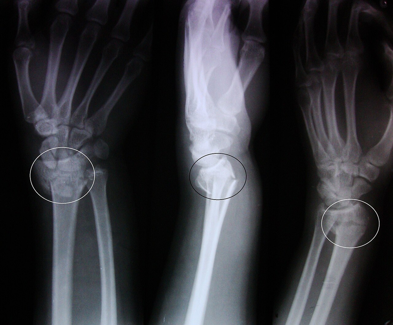
Photo: Ashish j29, CC BY 3.0
PART 3 · QUESTION 16
Model check
The model uses
Two real numbers for each site. Name them.
It leaves out
Name one thing about real people the model does not include.
Hint
Think about what makes one person’s bones different from another’s.
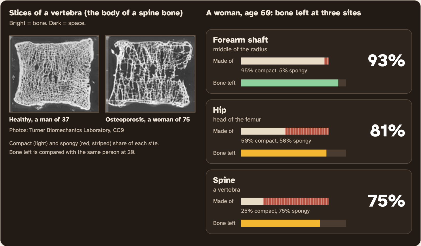
Screenshot: mrsajdak.com/bonetissue, Tab 3, a woman of 60
EXPLAIN IT · QUESTION 17
Why the hip, the spine and the wrist?
What I saw
One thing you measured or watched today.
The science idea
Where spongy bone is, and what osteoporosis does to it.
So
How that explains where the breaks happen.
A broken wrist: the end of the radius.
Photo: Ashish j29, CC BY 3.0
WRAP IT UP · BACK TO THE START
Revisiting the Phenomenon
Why is one bone built two ways: solid in the middle, full of holes at the ends?
1Using evidence from today, explain why the middle of the femur is a solid tube.
2Why is the end full of holes? Is it weaker, or just built differently?
3What lives in the holes in a living person?
4Which numbers from your worksheet best support your explanation?
Cut A: the shaft. Compact bone.
Cut B: near the hip. Spongy bone inside.
Photos: MAKY.OREL, Charles University, CC0 · Steven Fruitsmaak, public domain
LAST CHECK
Before you hand it in
Name, date and period are on top.
Every table is full.
Question 4 has one tick in each row.
Question 17 has all three parts.
Photo: OpenStax, CC BY 4.0