ANATOMY AND PHYSIOLOGY · CHAPTER 7 · THE LOWER LIMB
Bones of the Foot Lab
Follow along with your worksheet, one question at a time.
mrsajdak.com/foot
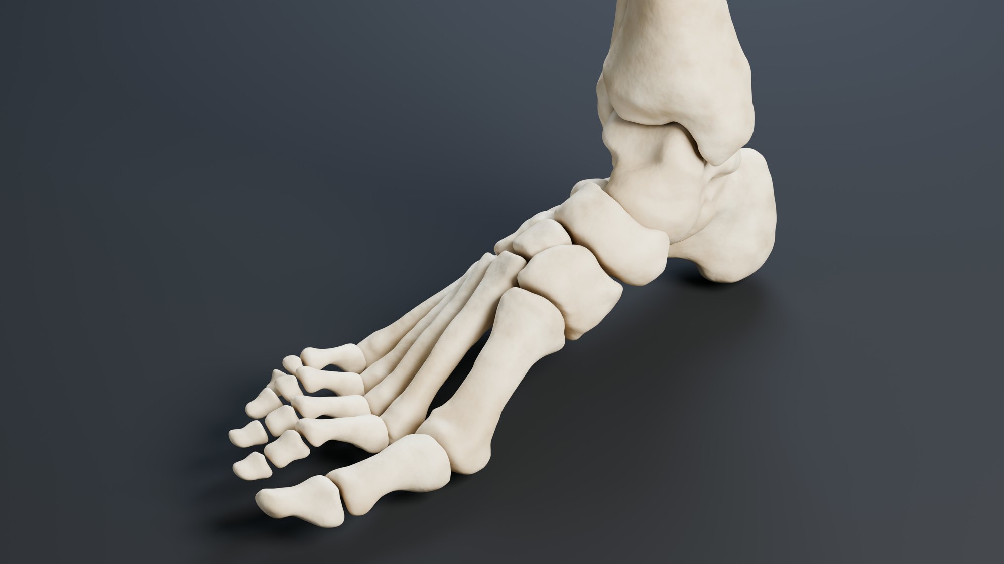
CT scan: Virtual Skeleton Database (SMIR), CC BY-NC-SA 4.0
START HERE · PICK AN OPENER
Choose how to open the lesson
Click a card to jump to it. Every option ends at the same Observe, Wonder, Predict slide and the same driving question.
OPTION A · VIDEO
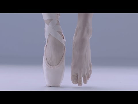
Feet at Work
Clip 1 is from The Australian Ballet: a close look at a dancer’s feet as she works. Clip 2 is a video X-ray of a living person’s ankle and big toe, moving. Clip 3 is a pressure mat: the colours show where a bare foot pushes on the floor.
Watch for
How many bones do you think are in one foot?
In the X-ray, which parts move? Which stay still?
On the pressure mat, which parts of the foot push hardest? Which part barely touches?
Videos on YouTube: The Australian Ballet; Arbor Foot Health Center; Tekscan Medical.
OPTION B · TRY IT · 5 MINUTES
The Paper Arch
1
Make two stacks of books about 15 cm apart.
2
Lay a sheet of paper flat across the gap like a bridge. Add coins to the middle one at a time. Count until it sags to the desk.
3
Now bend a second sheet into a curve, a hump, and wedge its ends between the book stacks so the ends cannot slide.
4
Add coins to the top of the arch, one at a time. Count until it gives way.
You need
Two sheets of printer paper per group
Four books (or two stacks of textbooks)
About 40 coins or metal washers of one kind
Watch for
Which held more: flat or arched?
What do the book stacks do for the arch? What happens if its ends slide out?
Where in your body could an arch help hold up weight?
OPTION C · PHOTOS
Phenomenon: Four Footprints
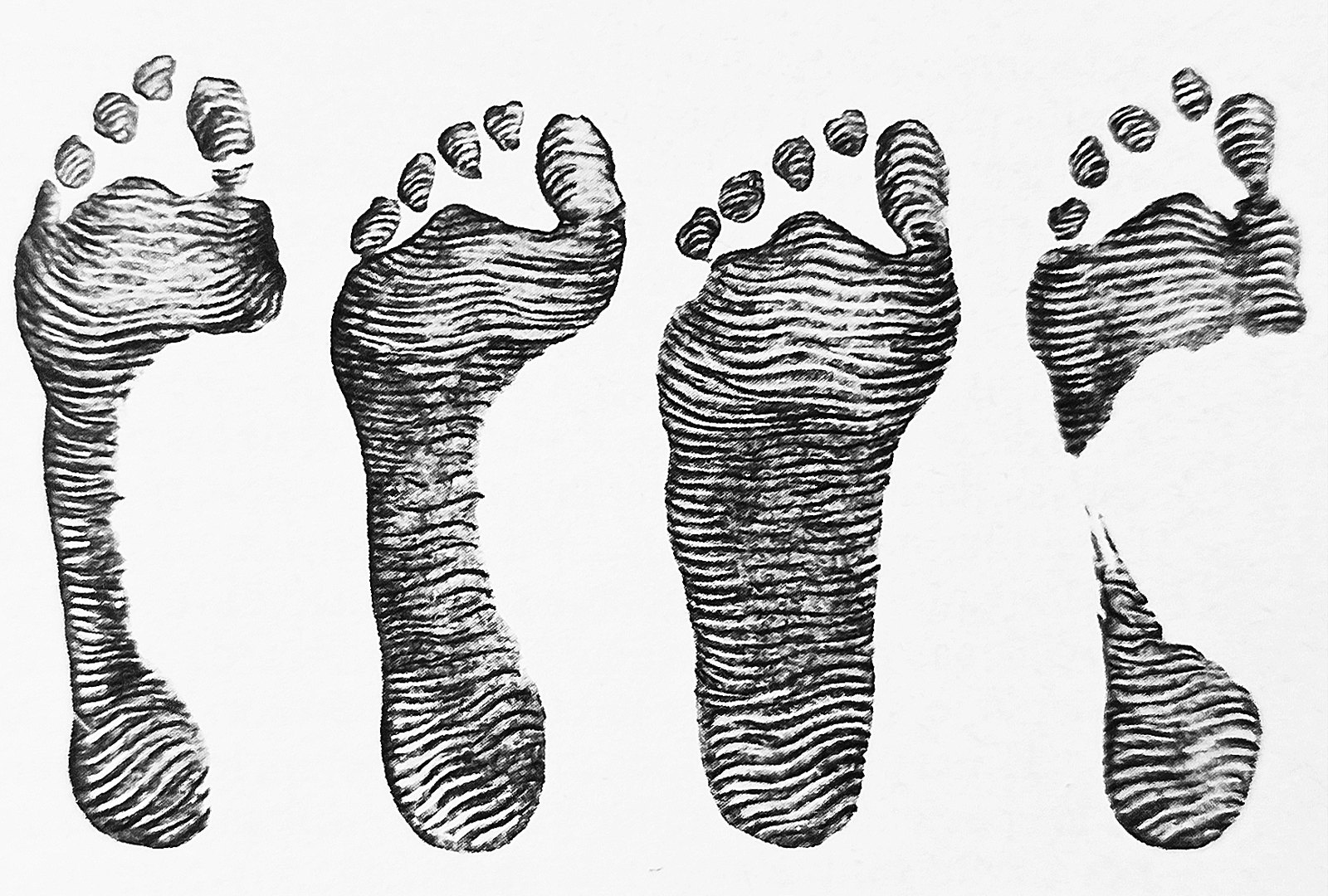
Footprints: Juan Carlos Fonseca Mata, CC BY-SA 4.0 · X-ray: Mikael Häggström, CC0
START HERE
Observe, wonder, predict
Every one of these people stands on the same kinds of bones.
OBSERVEIn Photo A, which parts of each foot touched the paper? Which did not?
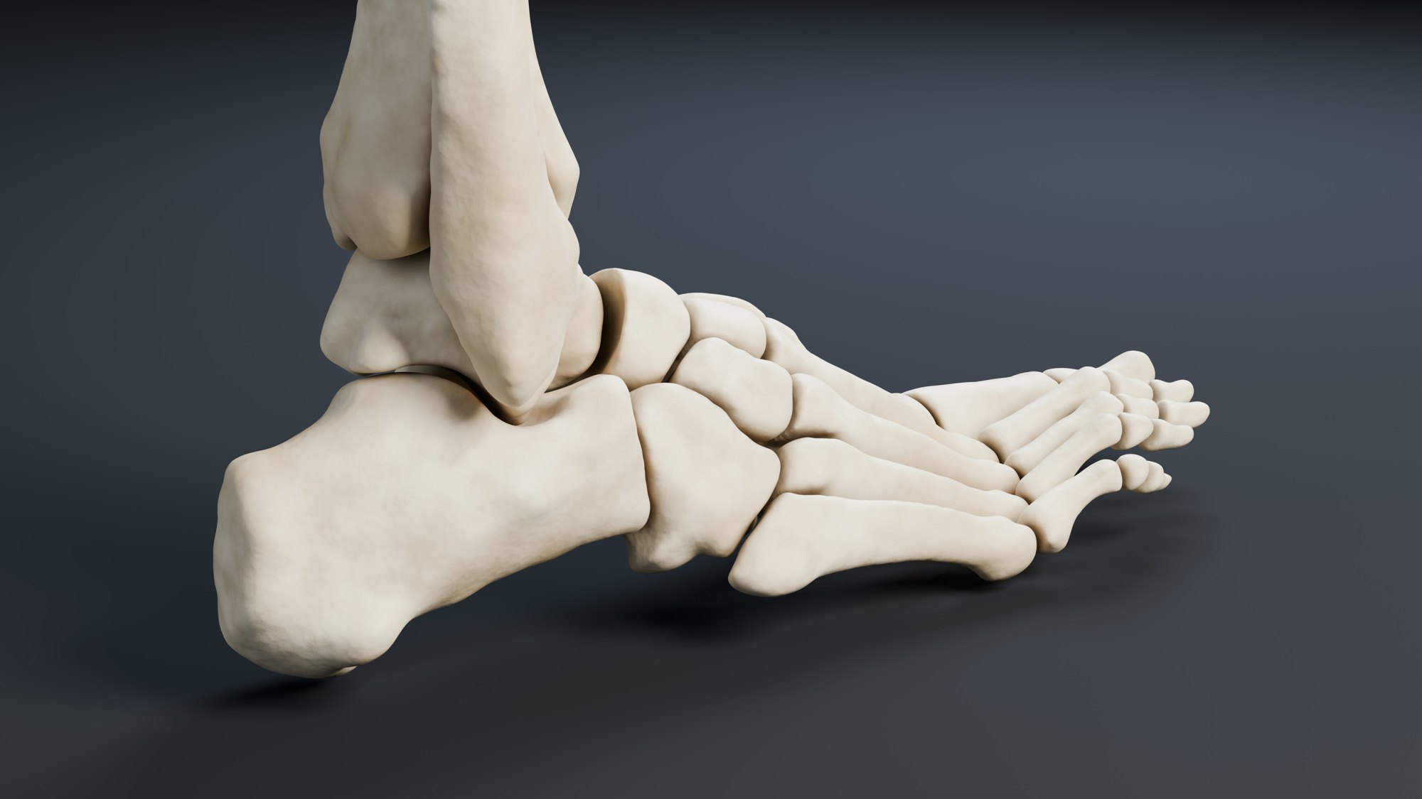
WONDERCount the bones you can see in Photo B. What surprises you?
PREDICTWhich bone in the foot do you think carries the most weight? Why that one?
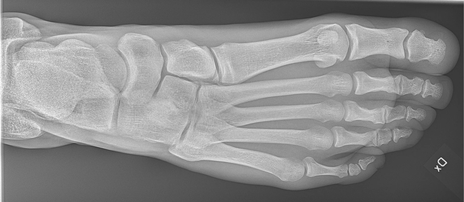
Photo B, up close.
Footprints: Juan Carlos Fonseca Mata, CC BY-SA 4.0 · X-ray: Mikael Häggström, CC0
DRIVING QUESTION
Your feet carry your whole body all day. How can 26 small bones hold up so much weight?
START HERE
What Happened Here?
Finished observing? Click here to find out.
The footprints come from four real people. Their feet have the same bones, but the bones sit at different heights, so different parts touch the floor. The X-ray is a normal right foot of a living person. The white shapes are bones, and there are more of them than most people guess.
Watched the video or tried it? Click here.
The dancer, the moving X-ray and the pressure mat all show a foot that is not one solid block. It is many small bones that move and share the load. The paper arch held more than the flat sheet. Why the foot's shape works so well is what this lab is about.
Today, you will name all 26 bones of a real foot from a CT scan, find the bone your whole leg rests on, and measure the arch that holds you up. At the end, you will come back to the driving question.
BEFORE YOU START
What you already know
From earlier lessons
An adult skeleton has 206 bones.
The lower leg has two bones: the thick tibia (shin) and the thin fibula.
A long bone is longer than it is wide. A short bone is about as wide as it is long.
Feel your own foot. How many separate bones can you find?
CT scan: Virtual Skeleton Database (SMIR), CC BY-NC-SA 4.0
BEFORE YOU START
What you will learn
I can locate and name the bones of the foot.
I can explain how the shape of the foot's bones fits their job of holding up the body.
BEFORE YOU START
Get set up
1
Open mrsajdak.com/foot on your device.
2
Write your name, the date and your period on the worksheet.
3
Find the tabs at the top of the lab. Each tab goes with one part of the worksheet.
4
Start on Tab 1. Do not skip ahead.

| Tab in the lab | Worksheet | Questions |
|---|---|---|
| 1. Name the 26 | Part 1 | 1 to 5 |
| 2. The ankle and heel | Part 2 | 6 to 10 |
| 3. The arch | Part 3 | 11 to 16 |
Screenshot: mrsajdak.com/foot
TAB 1 · QUESTIONS 1 TO 5
Part 1. Name the 26
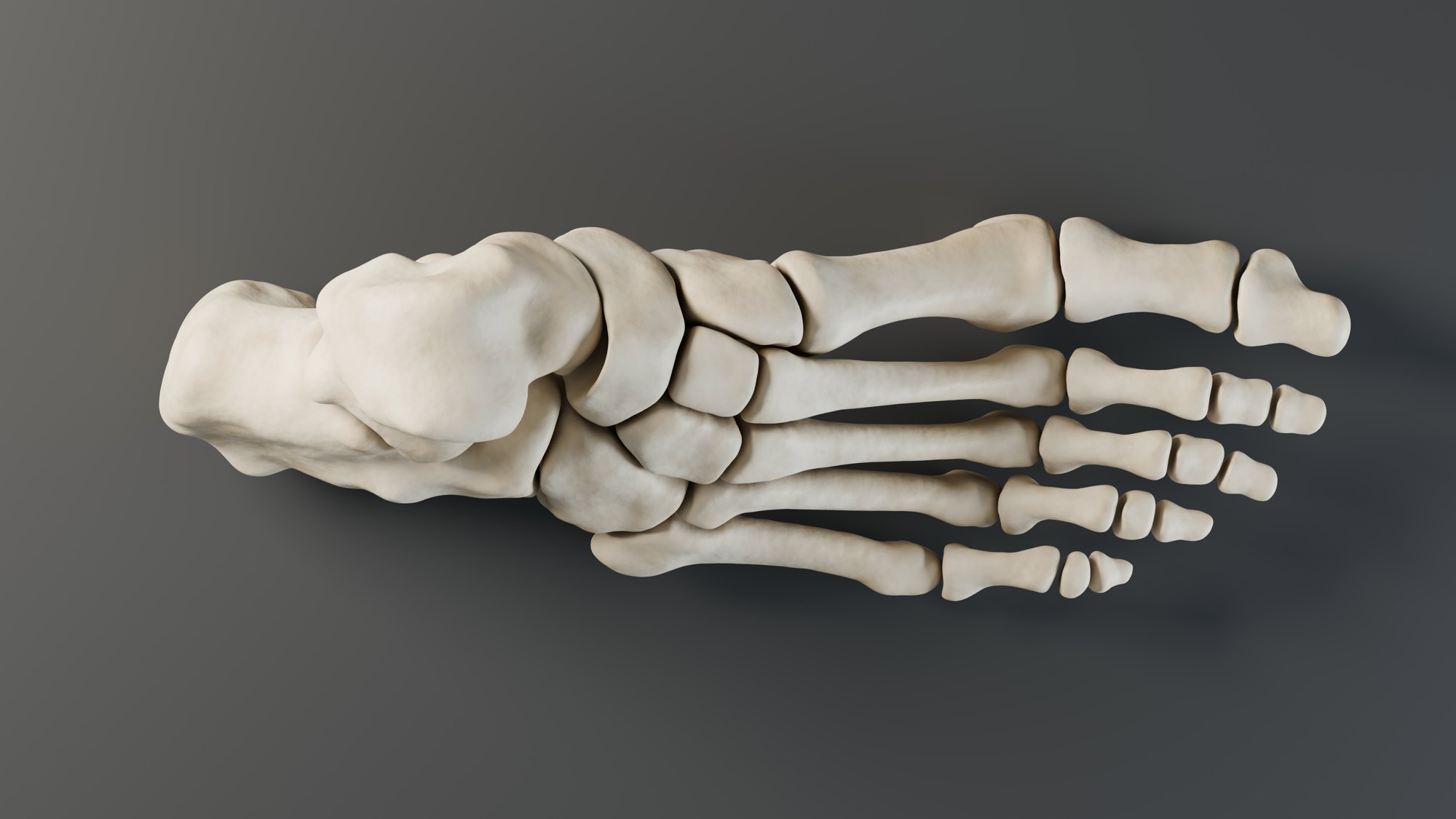
This is one man’s right foot, built from a CT scan.
Every bone is real, at full detail.
How many bones are there, and what are they called?
CT scan: Virtual Skeleton Database (SMIR), CC BY-NC-SA 4.0
PART 1 · QUESTIONS 1 AND 2
Find every bone
1
Click a bone on the photo. The lab names it.
2
The counts by group go up as you go.
3
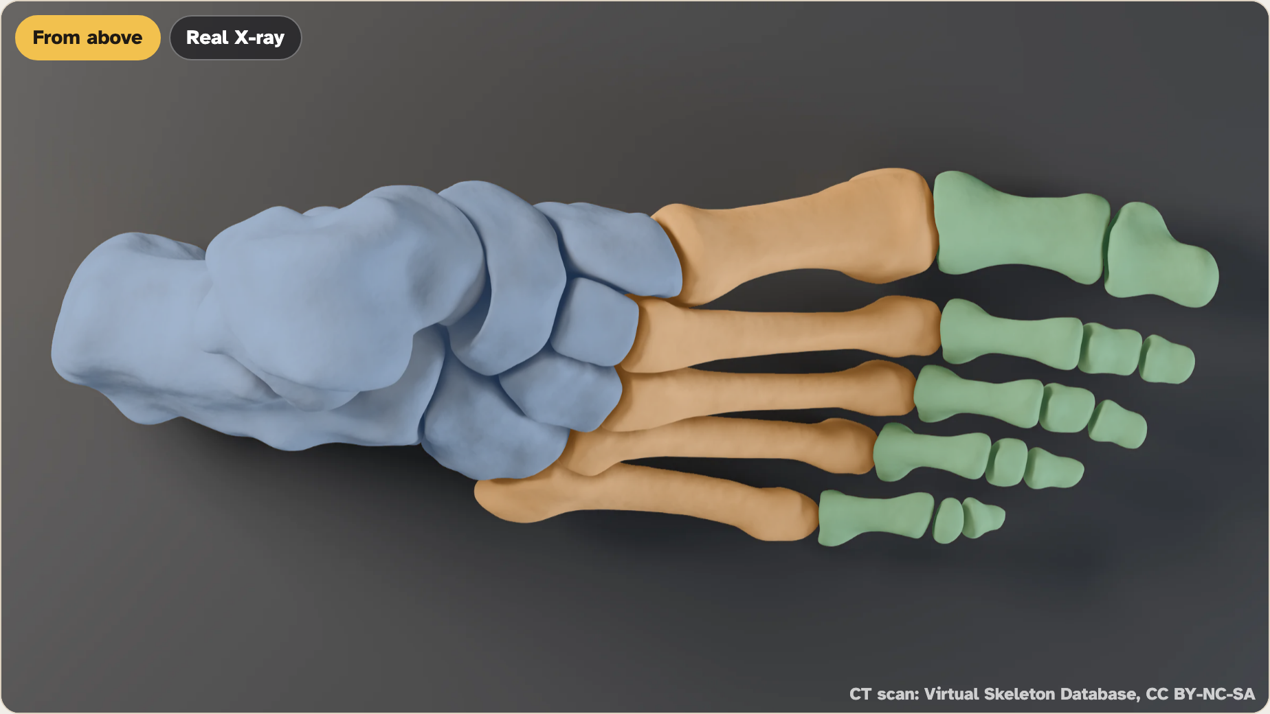
Turn on Color the groups.
4
Find all 26. Copy the counts from Your bones.
5
Click the toe bones one at a time and count them.
PART 1 · QUESTIONS 1 AND 2
Count by group
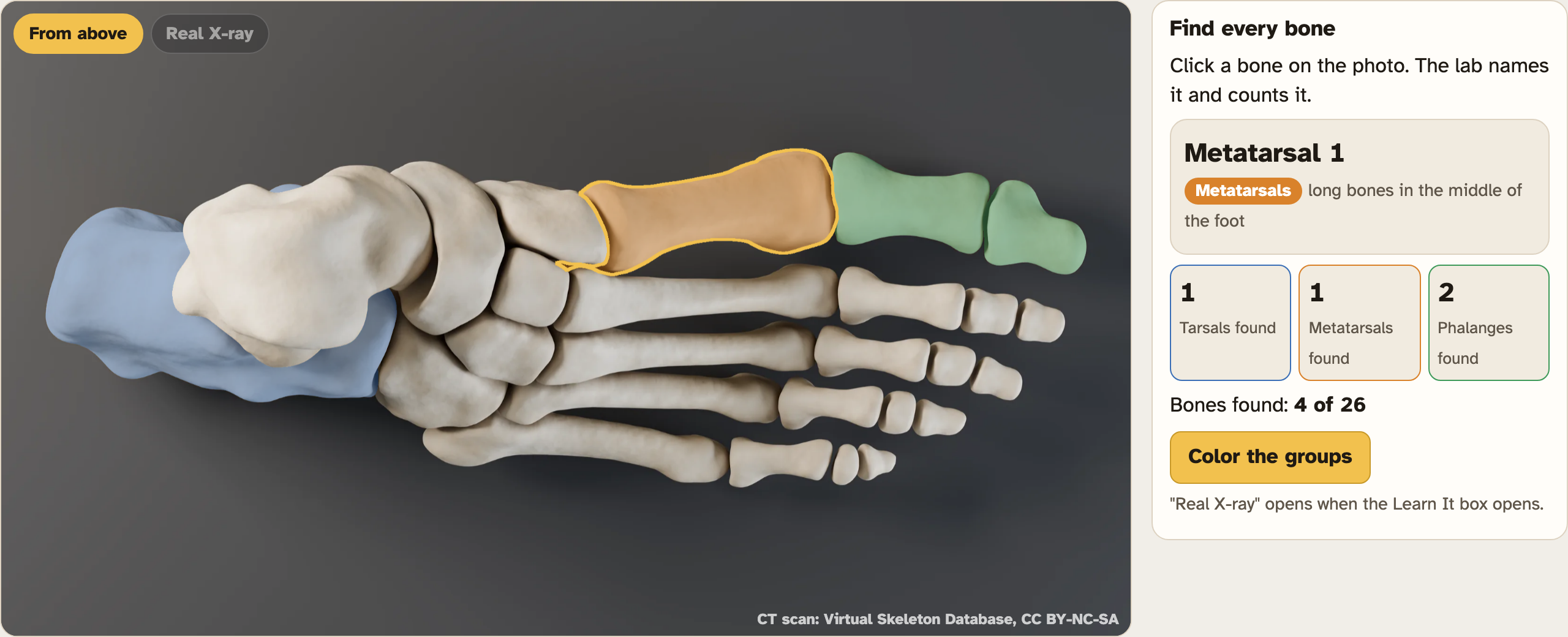
Four bones found so far.
QUESTION 1
How many in each group?
Find every bone, then copy the three counts and the total.
QUESTION 2
Count the toe bones
Does every toe have the same number of bones?
Screenshot: mrsajdak.com/foot, Tab 1 · CT scan: Virtual Skeleton Database (SMIR), CC BY-NC-SA 4.0
PART 1 · LEARN IT · 1 OF 4
Tarsals: 7 ankle bones
The back half of the foot is 7 tarsals. The calcaneus is the heel. The talus sits on top of it, under the shin. In front: the navicular, the cuboid and three cuneiforms.
Screenshot: mrsajdak.com/foot, Tab 1 · CT scan: Virtual Skeleton Database (SMIR), CC BY-NC-SA 4.0
PART 1 · LEARN IT · 2 OF 4
Metatarsals: 5 long bones
The middle of the foot has 5 metatarsals, numbered 1 to 5 from the big-toe side. Metatarsal 1 is short and thick.
Photo: CT scan: Virtual Skeleton Database (SMIR), CC BY-NC-SA 4.0
PART 1 · LEARN IT · 3 OF 4
Phalanges: 14 toe bones
Each small toe has 3 phalanges: proximal, middle and distal (the tip). The big toe, the hallux, has only 2. Your hand has the same plan: 8 + 5 + 14 = 27. Your foot: 7 + 5 + 14 = 26.
Photo: CT scan: Virtual Skeleton Database (SMIR), CC BY-NC-SA 4.0
PART 1 · LEARN IT · 4 OF 4
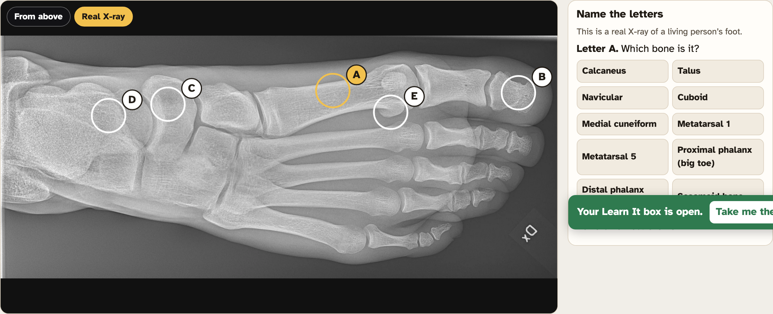
Extra little bones
Most people also have two pea-sized sesamoid bones under metatarsal 1, inside a tendon. They are not counted in the 26. The CT model leaves them out. A real X-ray shows them.
Screenshot: mrsajdak.com/foot, Tab 1 · CT scan: Virtual Skeleton Database (SMIR), CC BY-NC-SA 4.0
PART 1 · QUESTIONS 3 TO 5
Use the names
QUESTION 3
What share of you is feet?
Two feet, 206 bones in all.
Divide, then times 100.
QUESTION 4
Name by place
Which side is metatarsal 1 on?
What is the tip bone of a toe called?
Hand or foot: which has more?
QUESTION 5
A real X-ray
Press Real X-ray.
Name the bone at each letter. One letter is not in the 26.
PART 1 · THE BIG IDEA
The foot is a system of 26 bones in three groups: 7 tarsals, 5 metatarsals and 14 phalanges. It is built on the same plan as the hand.
IN YOUR WORDSHow is your foot built like your hand? How is it different?
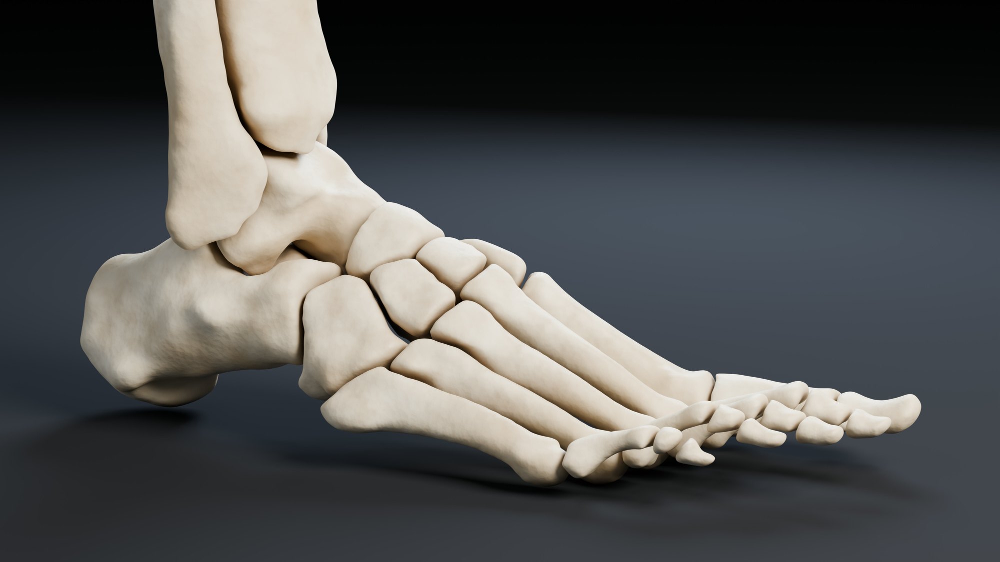
TAB 2 · QUESTIONS 6 TO 10
Part 2. The ankle and heel
Your shin comes down onto your foot.
But which foot bone does it actually rest on?
Turn the foot and find out.
CT scan: Virtual Skeleton Database (SMIR), CC BY-NC-SA 4.0
PART 2 · QUESTIONS 6 AND 7
Which bones touch?
1
Drag the photo left or right to turn the foot, or press Inside, Back, Outside or Front.
2
Click a bone. The lab shows its volume and the bones it touches.
3
Click the tibia, fibula, talus, calcaneus and navicular.
4
Copy each row from Your bones.
PART 2 · QUESTIONS 6 AND 7
Size and contact
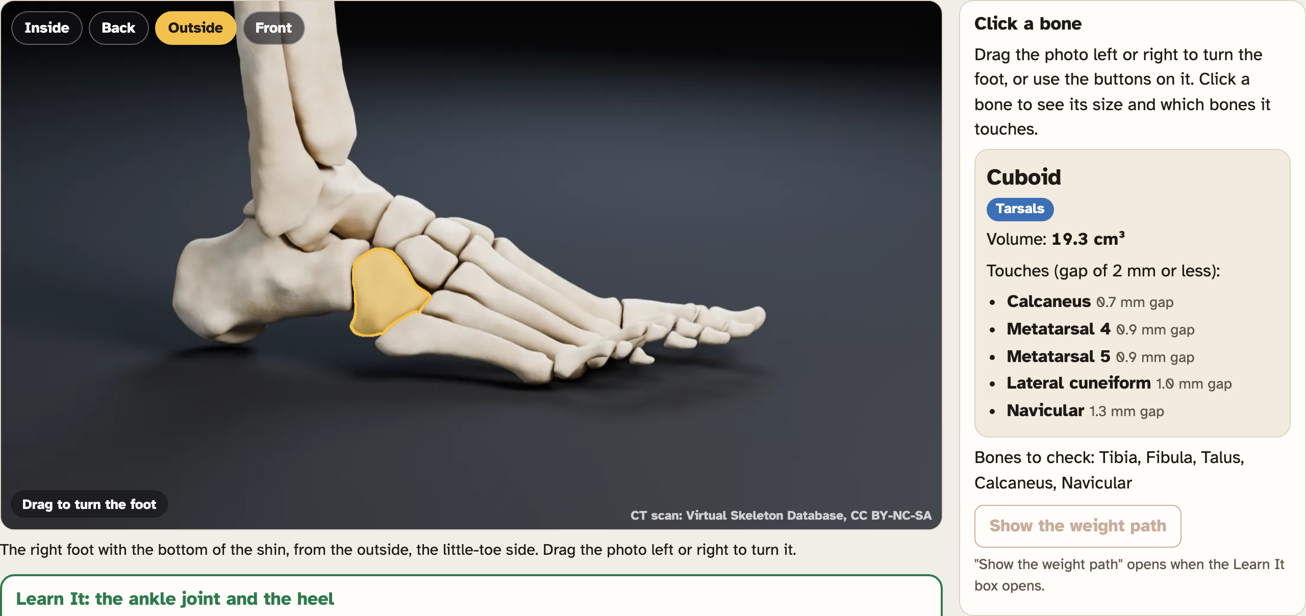
The cuboid, a bone not on your sheet.
QUESTION 6
Fill in the table
Volume and touches for five bones.
QUESTION 7
How many times bigger?
Calcaneus volume ÷ navicular volume.
Screenshot: mrsajdak.com/foot, Tab 2 · CT scan: Virtual Skeleton Database (SMIR), CC BY-NC-SA 4.0
PART 2 · LEARN IT · 1 OF 4
Where bones meet
A joint is where two bones meet. In this scan, bones at a joint sit 0 to 2 mm apart. In a living body, that gap is filled with smooth cartilage.
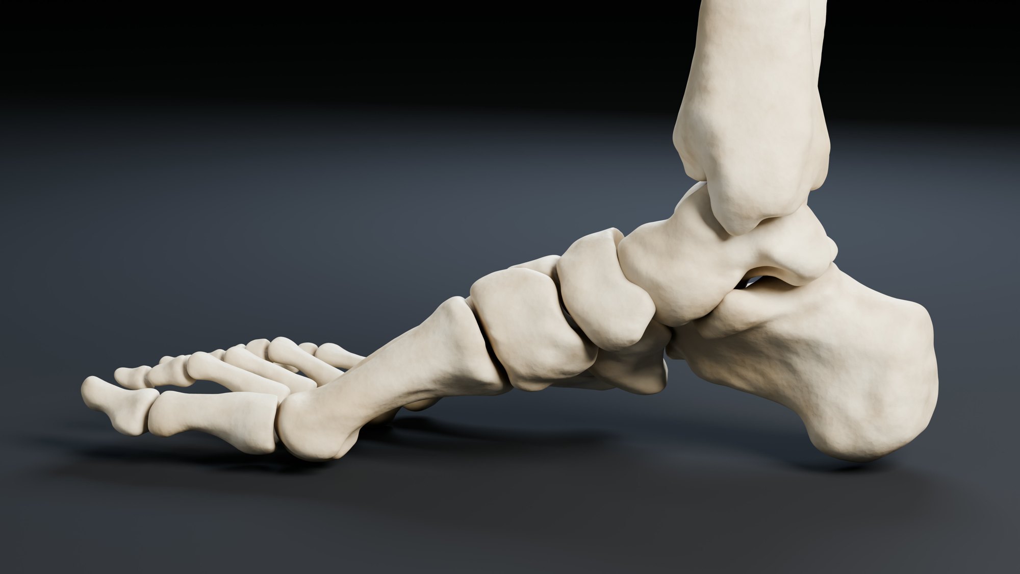
Photo: CT scan: Virtual Skeleton Database (SMIR), CC BY-NC-SA 4.0
PART 2 · LEARN IT · 2 OF 4
The ankle joint
The tibia and fibula grip the top of the talus, like a wrench around a bolt. The talus is the only foot bone the leg touches. All of your weight goes through it.
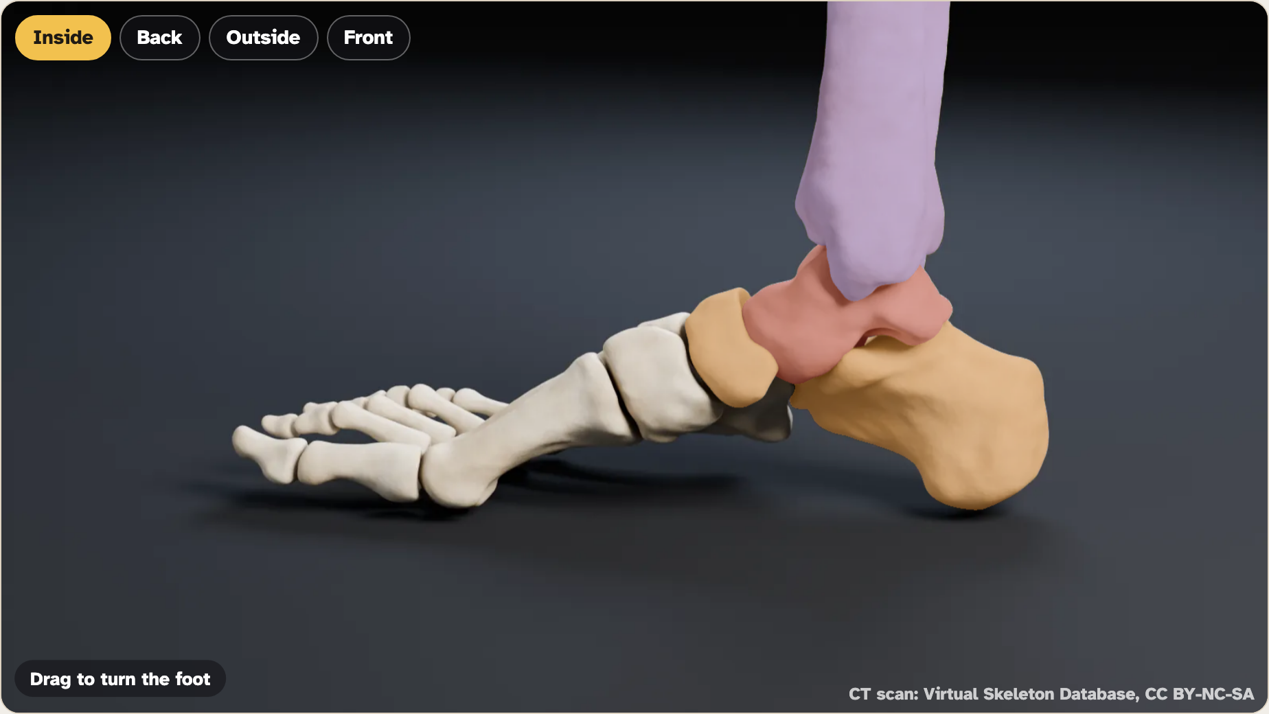
Screenshot: mrsajdak.com/foot, Tab 2 · CT scan: Virtual Skeleton Database (SMIR), CC BY-NC-SA 4.0
PART 2 · LEARN IT · 3 OF 4
The bumps on your ankle
The inside bump is the end of the tibia (medial malleolus). The outside bump is the end of the fibula (lateral malleolus).
Photo: CT scan: Virtual Skeleton Database (SMIR), CC BY-NC-SA 4.0
PART 2 · LEARN IT · 4 OF 4
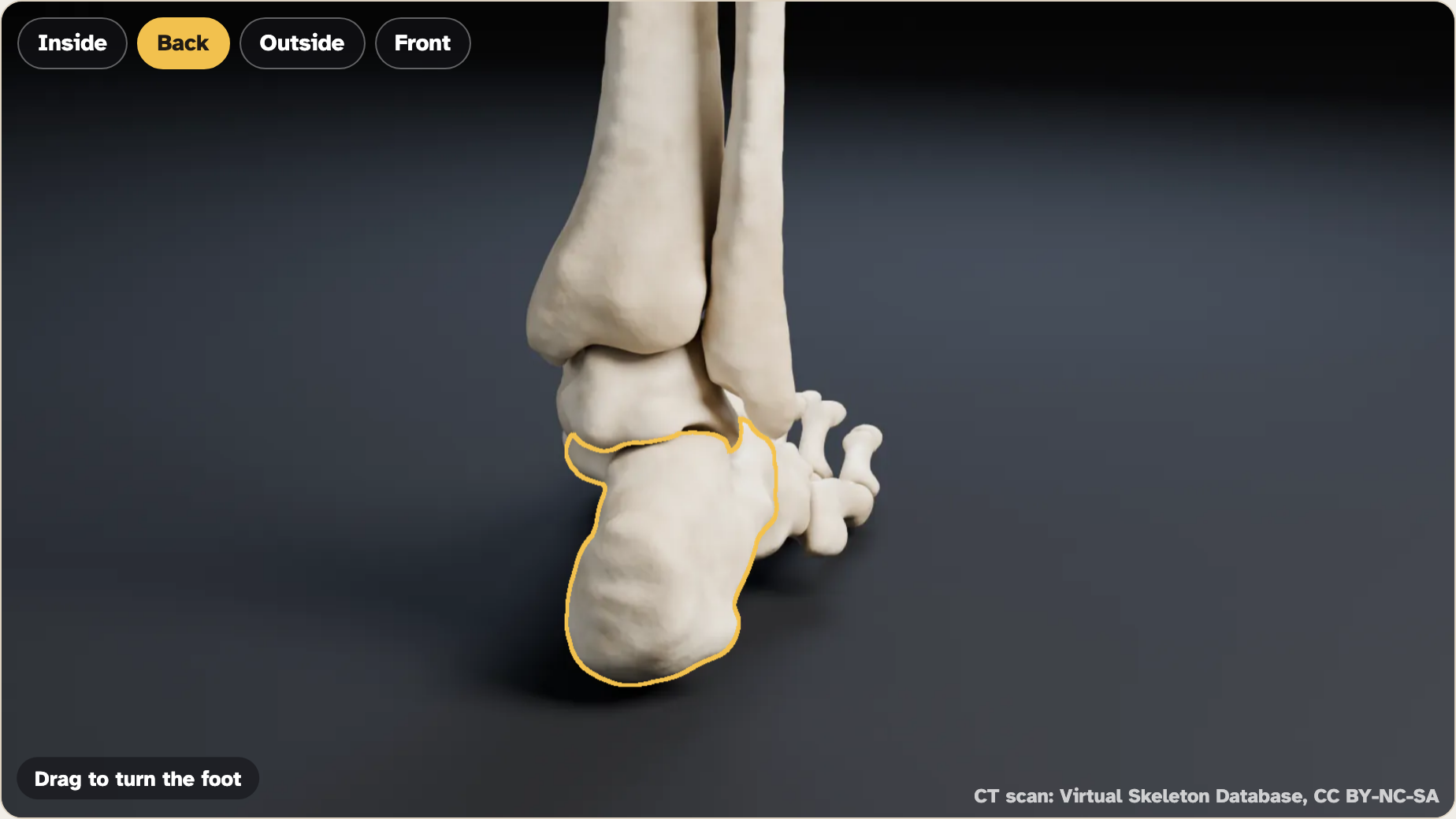
The biggest foot bone
From the talus, weight goes back into the calcaneus and forward to the toes. The calcaneus hits the ground first in each step. The Achilles tendon pulls on its back to lift your heel.
Screenshot: mrsajdak.com/foot, Tab 2 · CT scan: Virtual Skeleton Database (SMIR), CC BY-NC-SA 4.0
PART 2 · QUESTIONS 8 TO 10
Structure and function
QUESTION 8
Where the weight goes in
Which foot bone carries all of your weight from the leg?
Use your table to show how you know.
QUESTION 9
Two bumps
Inside ankle bump: which bone?
Outside ankle bump: which bone?
QUESTION 10
A big heel bone
Name one job that needs a big, strong heel bone.
PART 2 · THE BIG IDEA
The leg rests on one foot bone, the talus. It passes your weight back to the calcaneus and forward to the toes. The heel bone is the biggest because it takes the first hit and anchors the Achilles tendon.
IN YOUR WORDSWhy is the calcaneus so much bigger than the toe bones?
TAB 3 · QUESTIONS 11 TO 16
Part 3. The arch
Look at the inside of the foot.
Some bones touch the floor. Some do not.
How high is each one?
CT scan: Virtual Skeleton Database (SMIR), CC BY-NC-SA 4.0
PART 3 · QUESTIONS 11 AND 12
Measure the heights
1
Use Inside. Click the calcaneus, talus, navicular, medial cuneiform and metatarsal 1.
2
Use Outside. Click the cuboid.
3
Copy each height from Your heights.
4
Plot the five inside heights on your graph.
5
Press Show what touches the floor. Try From above too.
PART 3 · QUESTIONS 11 AND 12
Low or high?
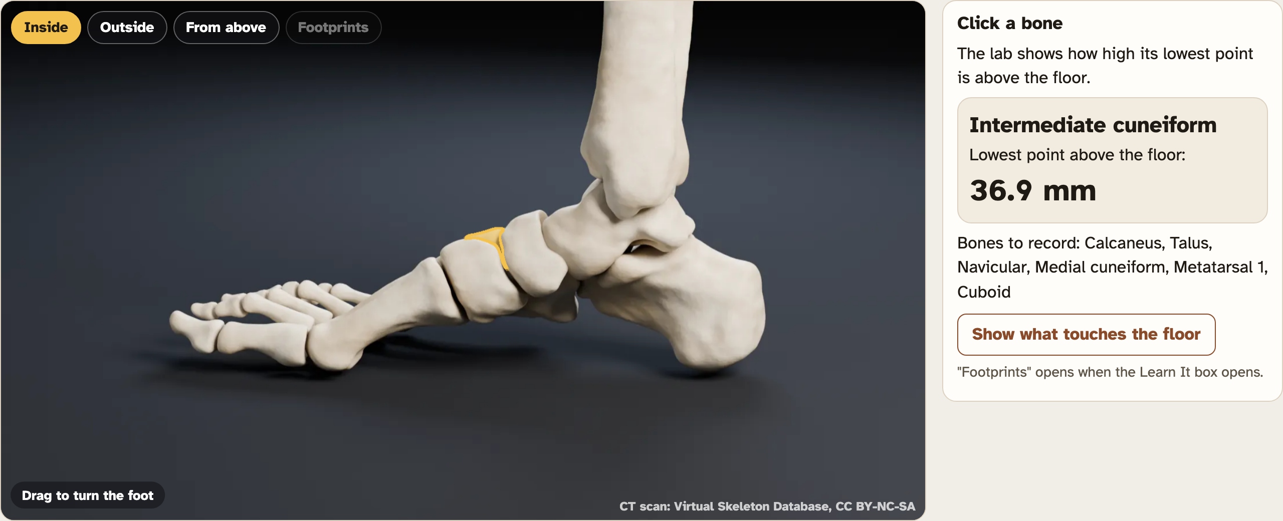
The intermediate cuneiform, a bone not on your sheet.
QUESTION 11
Copy the heights
Five inside bones, heel to toe, then the cuboid.
QUESTION 12
Plot them
Heel on the left. Connect the dots.
Which bones touch the floor?
Screenshot: mrsajdak.com/foot, Tab 3 · CT scan: Virtual Skeleton Database (SMIR), CC BY-NC-SA 4.0
PART 3 · LEARN IT · 1 OF 4
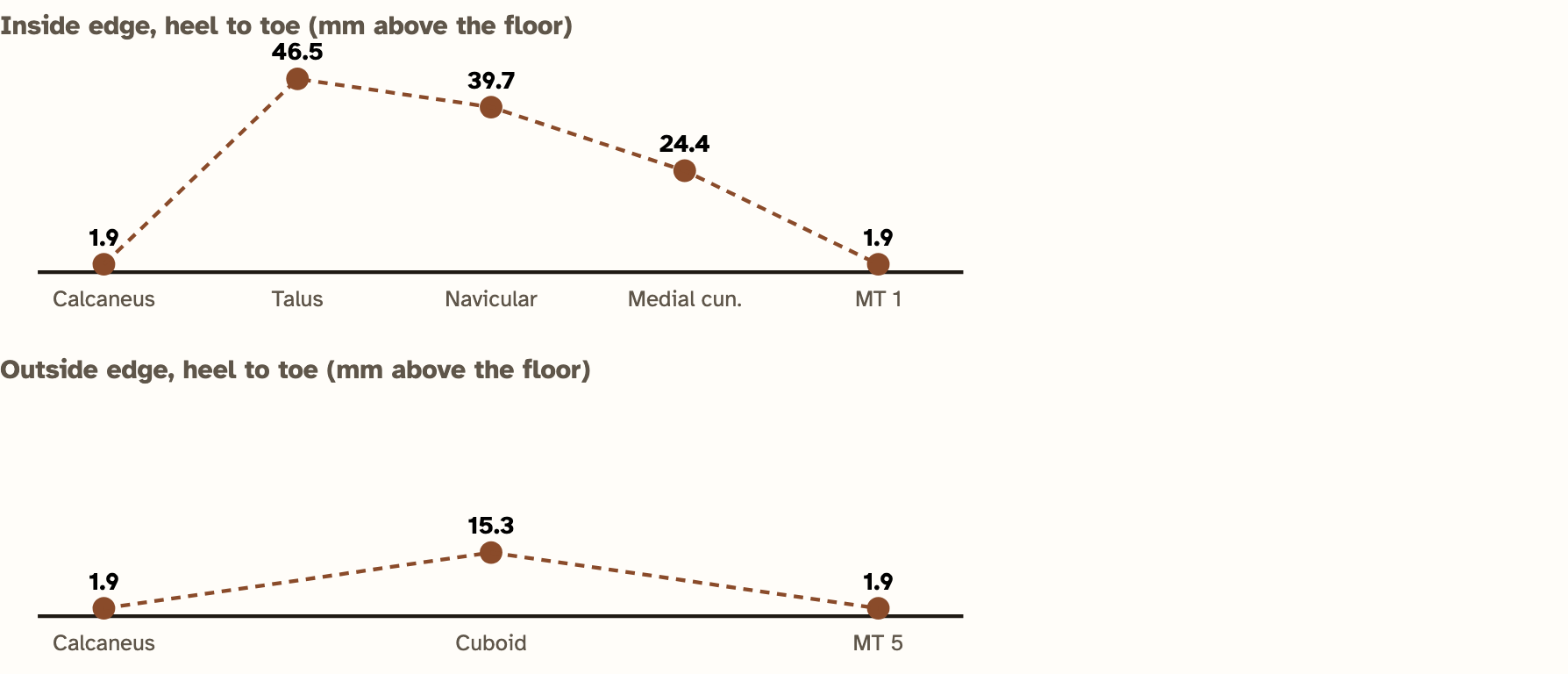
Low, high, low
Your heights went low, high, then low. That curve is an arch. This one runs along the inside of the foot: the medial longitudinal arch. The outside arch is lower.
Screenshot: mrsajdak.com/foot, Tab 3 · CT scan: Virtual Skeleton Database (SMIR), CC BY-NC-SA 4.0
PART 3 · LEARN IT · 2 OF 4
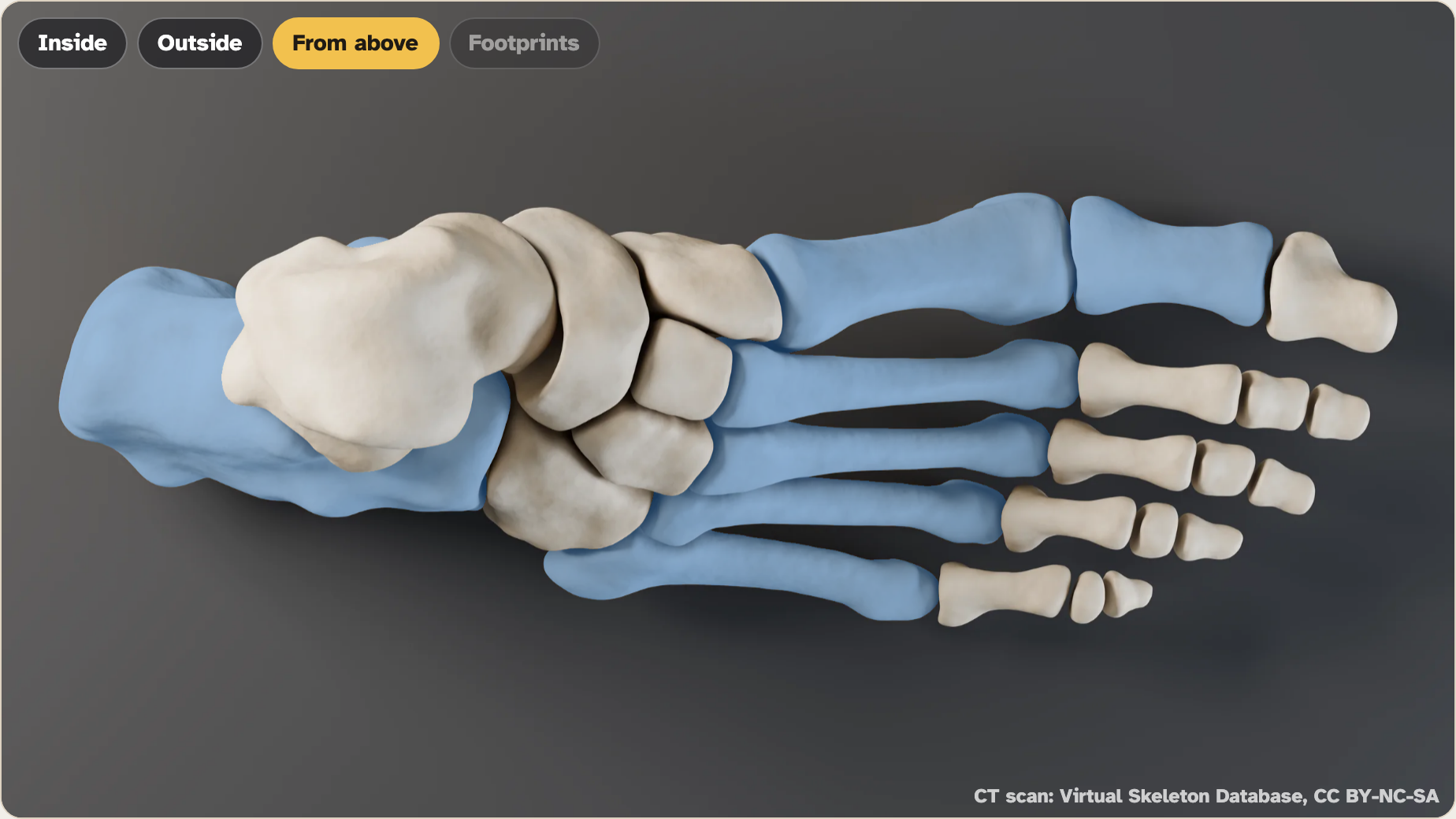
Why an arch?
An arch works like a stone bridge. It sends weight down to its two ends: the heel and the ball of the foot. Those are the bones that touch the floor. It also flattens a little and springs back with each step.
Screenshot: mrsajdak.com/foot, Tab 3 · CT scan: Virtual Skeleton Database (SMIR), CC BY-NC-SA 4.0
PART 3 · LEARN IT · 3 OF 4
What holds it up
Ligaments tie the bones together. A tough band under the sole, the plantar fascia, ties the heel to the toes like the string on a bow. Muscles help too. The model has bones only.
Photo: CT scan: Virtual Skeleton Database (SMIR), CC BY-NC-SA 4.0
PART 3 · LEARN IT · 4 OF 4
Flat feet and high arches
In a flat foot, the arch sinks. The navicular drops toward the floor and the footprint fills in. In a high arch, the footprint can break in two.
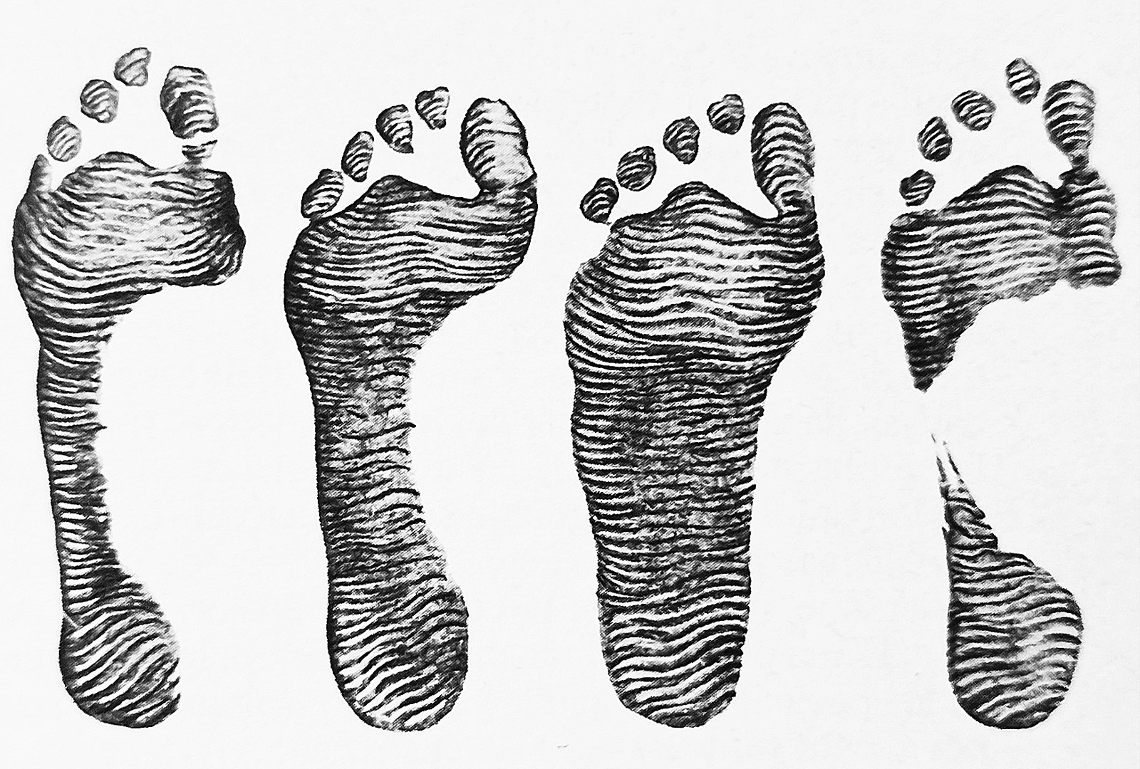
Photo: Juan Carlos Fonseca Mata, CC BY-SA 4.0
PART 3 · QUESTIONS 13 TO 15
Use the arch
QUESTION 13
Name the curve
What is the low, high, low shape called?
Which two numbers show the inside arch is higher?
QUESTION 14
Judge the footprints
Press Footprints. Which print came from the flattest foot?
What happens to the navicular?
QUESTION 15
Model check
What does the model leave out that holds the arch up?
What does letter E on the X-ray show that the model misses?
PART 3 · QUESTION 16
Explain it
Question 16
How can 26 small bones hold up your whole body?
What happens
Where your weight goes into the foot and down to the floor.
The science idea
How the bones’ shapes and the arch fit the job.
How the evidence shows it
Use two numbers from this lab.
PART 3 · THE BIG IDEA
The foot’s bones form arches. An arch sends your weight to its two ends, the heel and the ball of the foot, and springs a little with each step. Ligaments and the plantar fascia hold it in shape.
IN YOUR WORDSWhy does a flat foot leave a filled-in footprint?
WRAP IT UP · BACK TO THE START
Revisiting the Phenomenon
Your feet carry your whole body all day. How can 26 small bones hold up so much weight?
1Go back to the footprints. Explain the gap in the far-left print using the bones.
2Your PREDICT answer: which bone carries the most weight? Were you right? What would you change?
3Name one bone in each group: a tarsal, a metatarsal and a phalanx. Where is each one?
4A friend says the foot would be stronger as one solid bone. What would it lose?
Footprints: Juan Carlos Fonseca Mata, CC BY-SA 4.0 · X-ray: Mikael Häggström, CC0
LAST CHECK
Before you hand it in
Name, date and period are on top.
Both sides are done.
Every table is full and your graph is drawn.
Your explanation uses two numbers from the lab.
CT scan: Virtual Skeleton Database (SMIR), CC BY-NC-SA 4.0